Figure 4. Immunohistochemistry staining for COL1A2 (left), RAB22A (middle), and negative control (primary antibody omitted; right) in
sagittal sections of paraffin- embedded basset hound (BH) eyes derived from affected and unaffected animals. Variable intensities
of COL1A2 and RAB22A staining were noted in the A: ciliary body (CB) and processes, B: trabecular meshwork (TM) and ciliary cleft (CC), C: stroma and pigmentary epithelium (PE) layers of the iris, D: epithelium (EP), stroma and endothelium (ED) layers of the cornea, E: retina (OS=outer segment, ONL=outer nuclear layer, OPL=outer plexiform layer, INL=inner nuclear layer, IPL=inner plexiform
layer, GCL=ganglion cell layer; E), and F: optic nerve axons (ONA). Arrows indicate areas where staining was prominently observed.
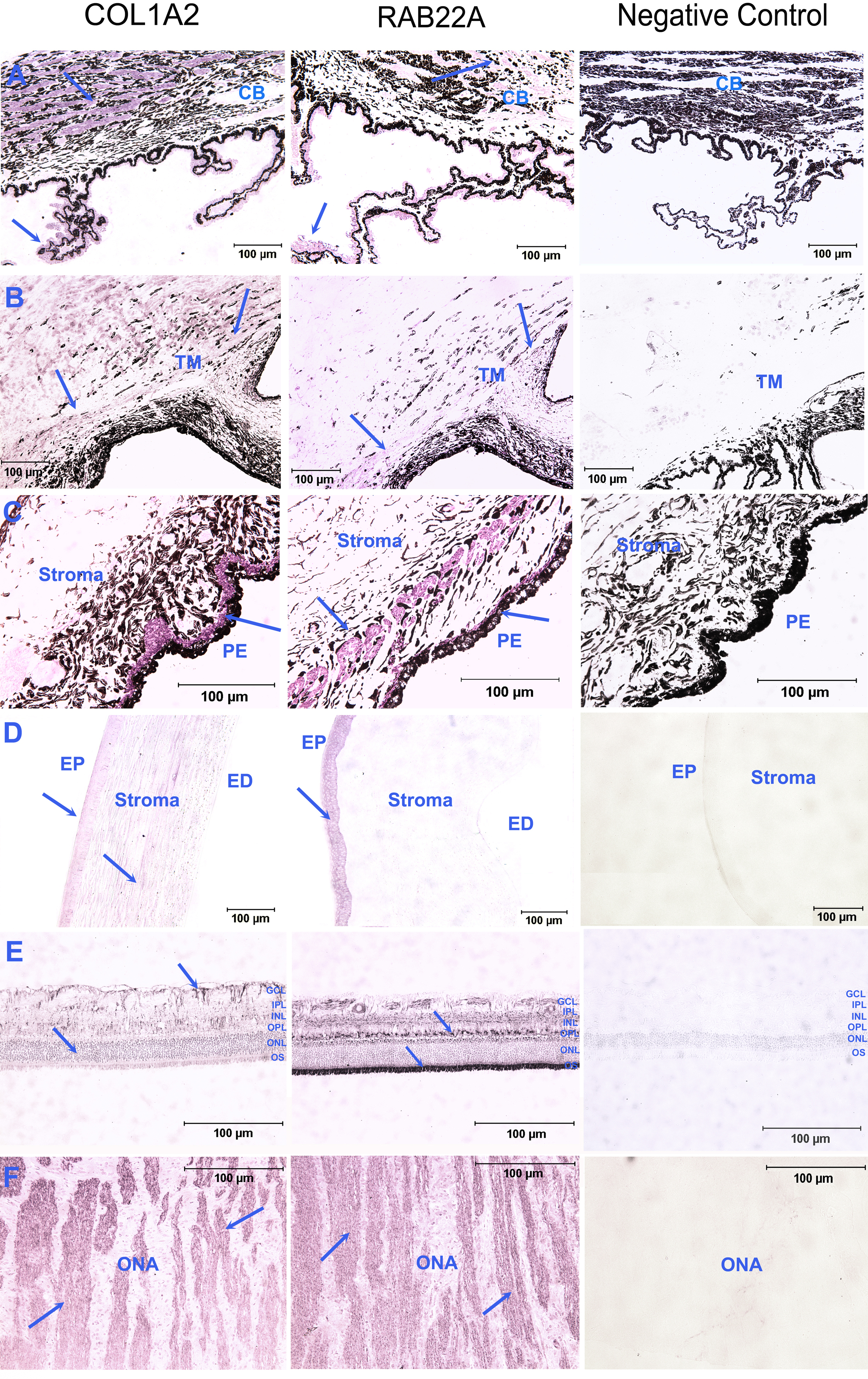
 Figure 4 of
Ahram, Mol Vis 2014; 20:497-510.
Figure 4 of
Ahram, Mol Vis 2014; 20:497-510.