Figure 2. Analysis of productive Cre-mediated recombination by examining the frequency of β-galactosidase (β-gal)-expressing RPE cells
in F1 mice derived from inducible RPE-specific Cre and ROSA26 lacZ reporter mice. A–D: Representative images and statistical analysis of immunostained RPE flatmounts for Cre-activated β-gal (green). A: An enlarged confocal image shows the number and frequency of β-gal-expressing RPE cells. Scale bar = 40 µm. B–C: Representative images show the number and frequency of β-gal-positive cells in central (B) and peripheral RPE flatmounts (C). Scale bars = 100 nm. D: Statistical analysis with the Student t test shows that there was no significant difference in the frequency of β-gal-positive RPE cells between the central and
peripheral regions of the RPE. Error bar: standard deviation (SD), n = 5–6. E: Representative image shows homogenous Cre-activated β-gal activity in the retinal section of the Cre/β-gal double transgenic
mice. Scale bar = 100 µm. Productive-Cre mediated recombination occurred evenly in approximately 60% of the RPE cells in inducible
RPE-specific Cre mice after a single intravitreal doxycycline (Dox) injection.
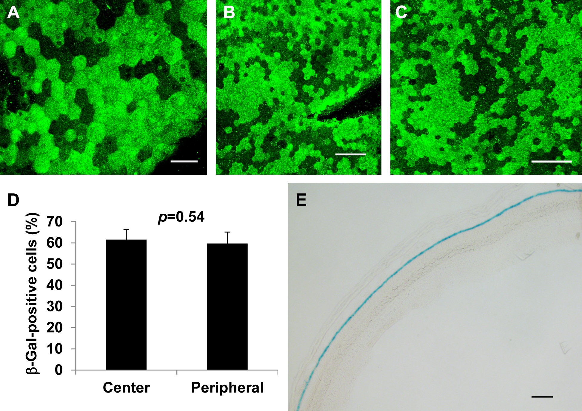
 Figure 2 of
Fu, Mol Vis 2014; 20:480-487.
Figure 2 of
Fu, Mol Vis 2014; 20:480-487.