Figure 4. Immunoconfocal microscopy for keratin 13 (A–D), 19, (E–H), MUC5AC (I–L), and connexin 43 (M–P). In patients 1–3, staining for keratin 13 is unanimously negative (A–C); however, in Patient 4, strong suprabasal staining for keratin 13 is evident in the central cornea but not in the peripheral
cornea (D). Full-thickness epithelial staining for keratin 19 is seen in Patients 1 and 4 (E, H), and the staining is predominantly in the basal as well as the suprabasal epithelium in Patients 2 and 3 (F, G). In patients 1 to 4, the signal for MUC5AC is universally negative, suggesting that none contains goblet cells (I–L). The insert in 4L shows normal conjunctiva containing goblet cells. In Patients 1 to 3, connexin 43 (Cx43) staining is present in the entire
layer of the epithelium (M–O). However, Cx43 staining is predominantly in the superficial and suprabasal epithelial layer in Patient 4 (P).
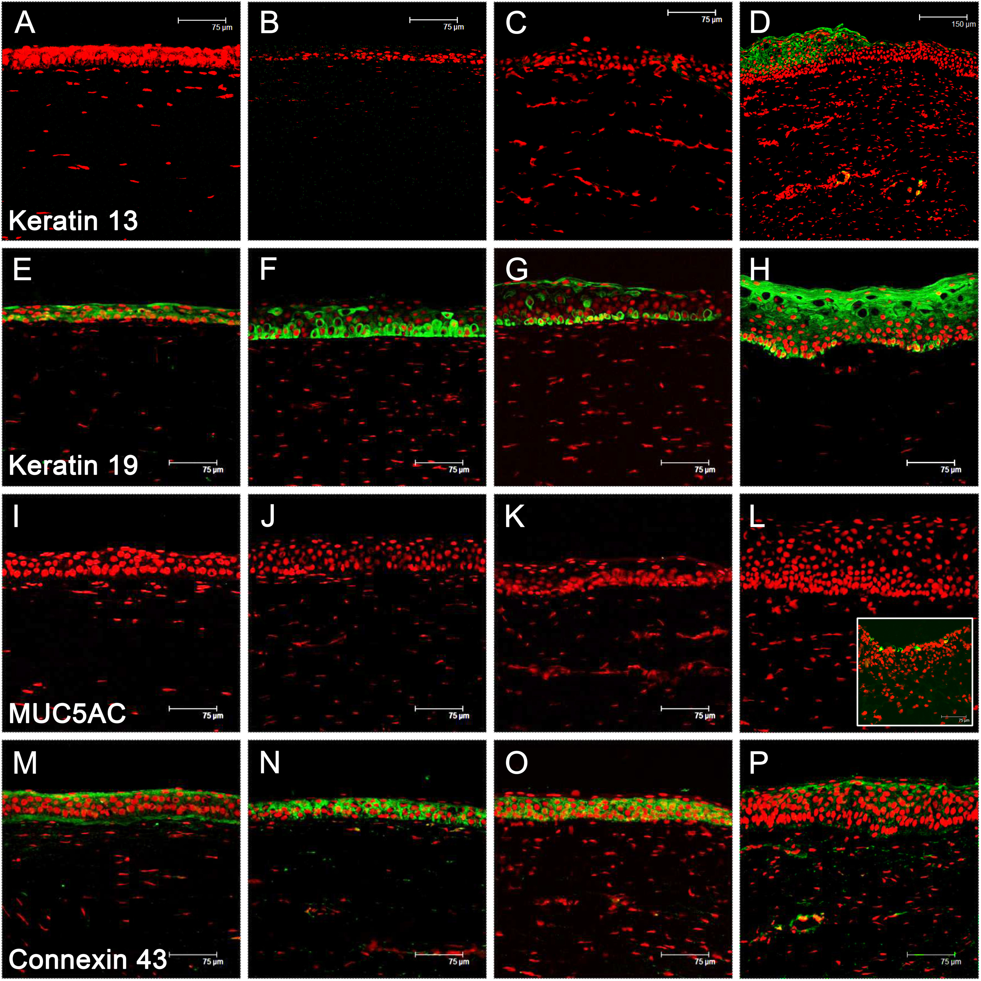
 Figure 4 of
Ma, Mol Vis 2014; 20:468-479.
Figure 4 of
Ma, Mol Vis 2014; 20:468-479.