Figure 3. Immunoconfocal microscopy for keratin 3 (A–D) and 12 (E–H) in Patients 1–4, and double staining of keratin 12 (green) and 13 (red) in Patient 4 (I). In all patients, homogenous keratin 3 can be seen; however, the signal for keratin 3 was negative in the basal epithelial
layer in Patient 3 (C) and the central cornea of Patient 4 (D insert). Keratin 12 is expressed homogenously in Patients 1 and 2 (E, F), but is negative in some basal epithelial cells in Patient 3 (G). In Patient 4, keratin 12 is positive in the peripheral cornea (H, I), but entirely negative in the central cornea except in focal areas (I, insert). In Patient 4, the central cornea is keratin 13 positive, suggesting the invasion of the conjunctival epithelium
(I). This pattern is due to vertical invasion of the conjunctival epithelium, but the corneal button was bisected horizontally.
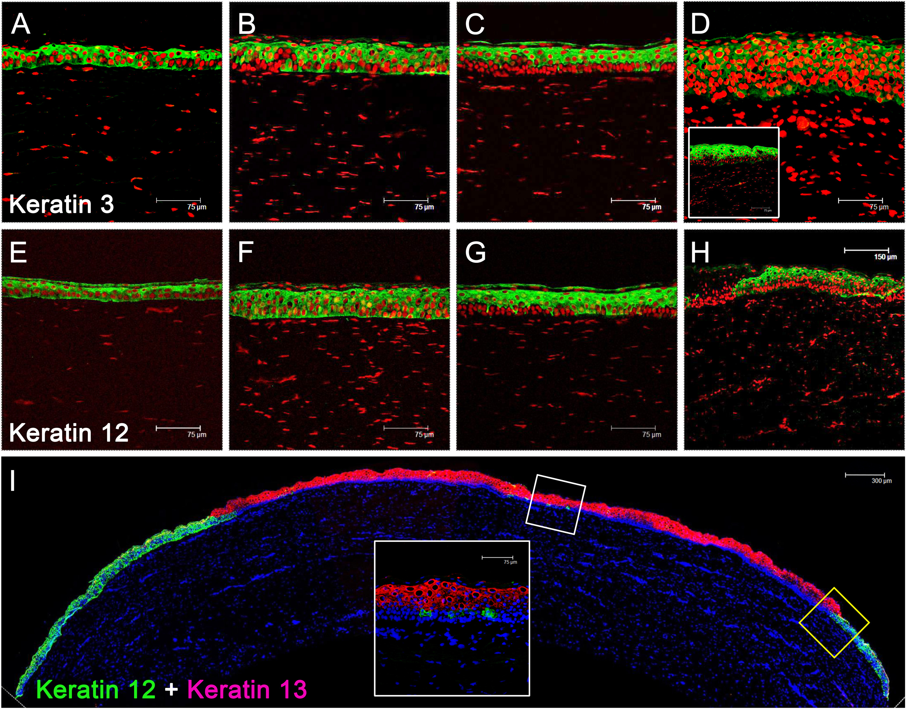
 Figure 3 of
Ma, Mol Vis 2014; 20:468-479.
Figure 3 of
Ma, Mol Vis 2014; 20:468-479.