Figure 2. Transmission electron microscopy of normal cornea (A–D) and corneal specimens from Patients 2 (E–H) and 3 (I–L). In the normal cornea, collagen fibrils in the stroma are uniformly organized in a lamellar arrangement (A). Regular arrangement of collagen fibrils in a longitudinal view (B) and a cross-sectional view (C), with an average fiber diameter of 35 nm. Bowman’s membrane (arrowhead) is present in the normal cornea (D). Irregular and variably thinned collagen lamellar layers are seen inP 2 (E) and 3 (I), respectively. Disorganized, nonlinear, and random distribution of collagen fibrils in the corneal stroma is also found
(F, G, J, and K). The collagen fibers varied considerably in diameter from 46 to 78 nm. In Patients 2 and 3, Bowman’s membrane is nearly
invisible (H, L). In contrast to the normal cornea in which the anterior stromal collagen is loosely organized (D), the collagen fibers in the anterior stroma of Patient 2 are more densely arranged (H).
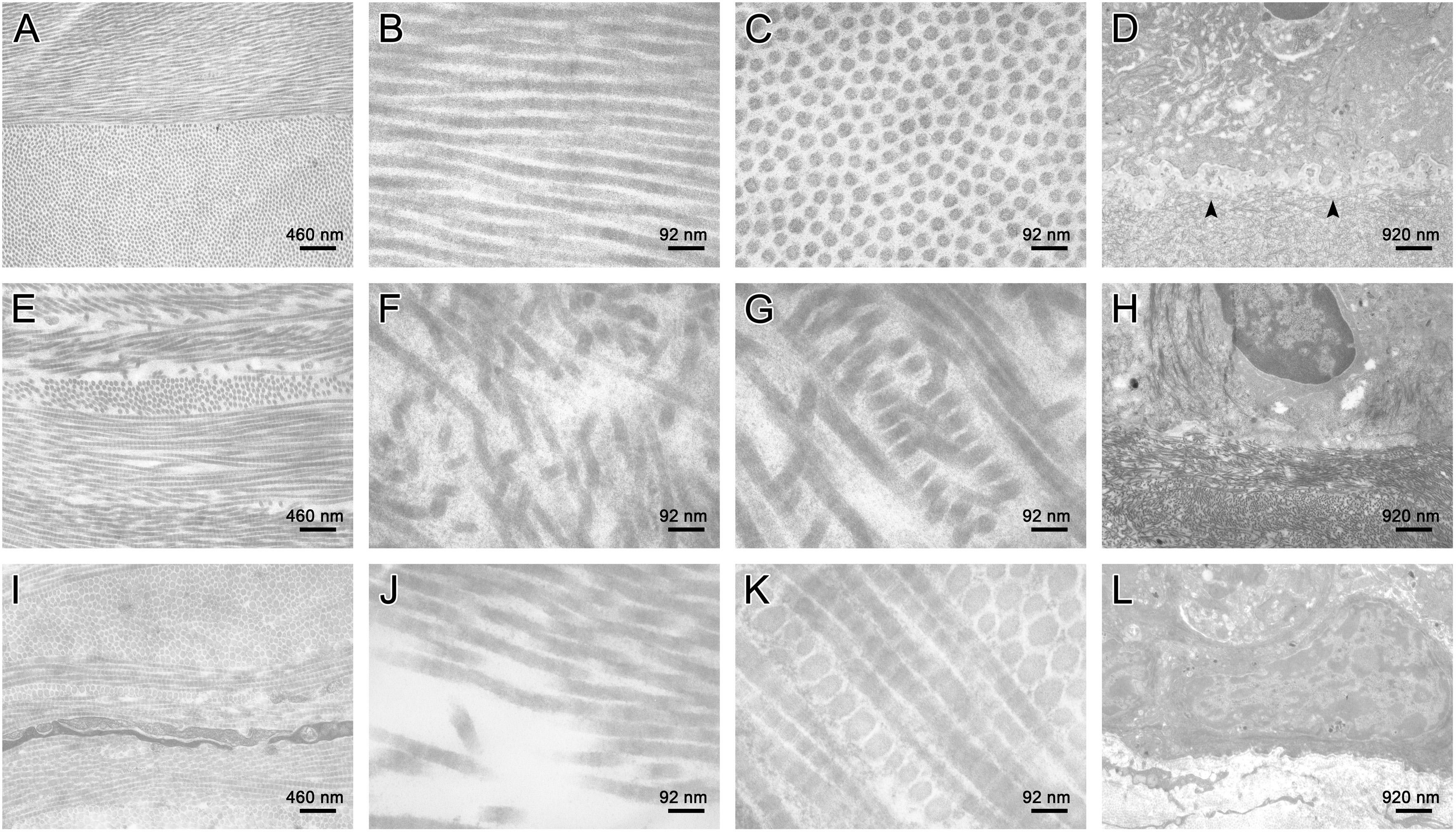
 Figure 2 of
Ma, Mol Vis 2014; 20:468-479.
Figure 2 of
Ma, Mol Vis 2014; 20:468-479.