Figure 1. External eye photos and hematoxylin-eosin staining of corneal specimens from four patients with total sclerocornea. A–H: Preoperative photos of Patients 1–4. I–P: Photos taken at postop 25, 32, 20, and 12 months, respectively. Q–T: Hematoxylin-eosin (HE) staining of corneal specimens viewed at 40X. U–X: Same specimens viewed at 400X, with emphasis on the corneal epithelium. Note that prominent corneal neovascularization from
5 to 8 O/C is seen in Patient 4 (H). Arrowhead: blood vessel in corneal stroma; arrow: iris pigment adhered to posterior corneal stroma; arrow in X: junction
between corneal (left) and conjunctival epithelium (right) in Patient 4.
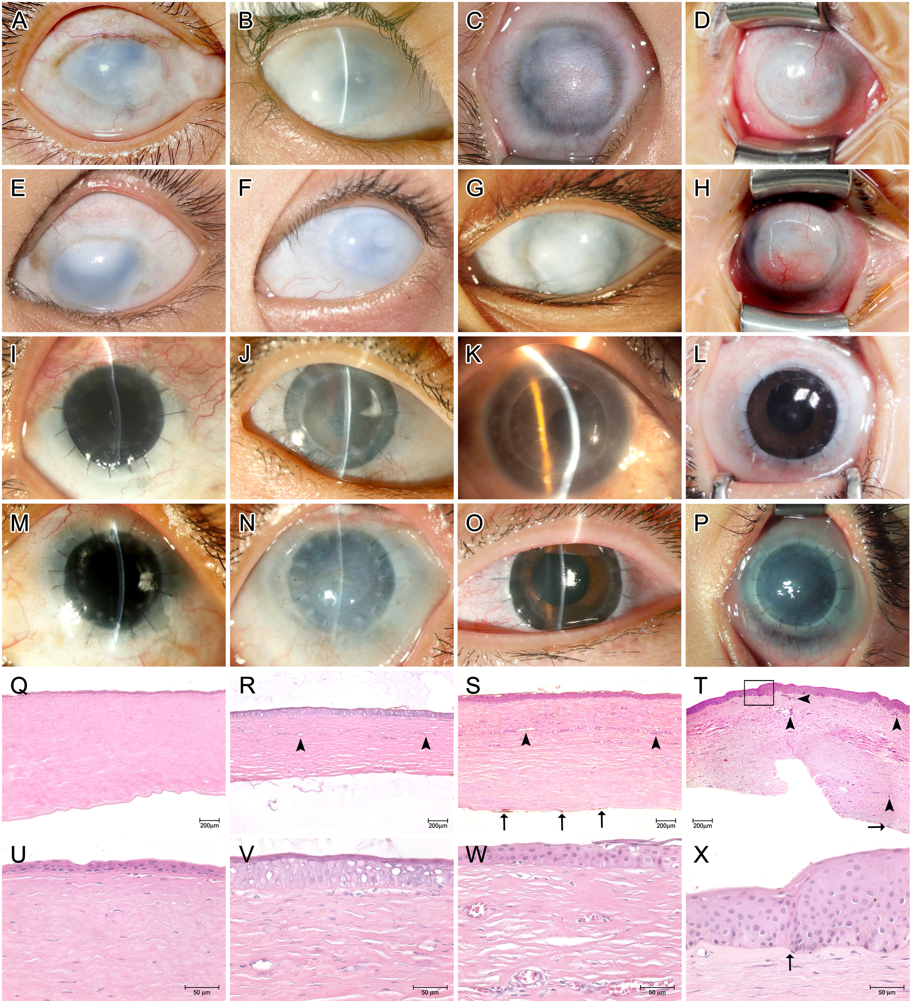
 Figure 1 of
Ma, Mol Vis 2014; 20:468-479.
Figure 1 of
Ma, Mol Vis 2014; 20:468-479.