Figure 2. Diphenyleneiodonium (DPI) inhibitor markedly attenuated the hydrogen peroxide (H2O2) generated signal in lens cells detected by peroxyfluor-6 acetoxymethyl ester (PF6-AM). A, top panel: Model indicating (red box) the region of E10 chick embryonic lens containing the equatorial zone (EQ) that is
imaged below. A, bottom left panel: Lens placed in organ culture incubated with PF6-AM (green) showing H2O2 production in the EQ zone. A, bottom right panel: Lens placed in organ culture incubated with PF6-AM (green) before exposure to the inhibitor diphenyleneiodonium
(DPI) attenuates PF6-AM fluorescence, suggesting the presence of an endogenous H2O2 signal. Both images are shown as an overlay with the differential interference contrast (DIC) image. B: Primary cultures of lens epithelial cells. (Top panels) Cultured lens epithelial cells incubated with PF6-AM (green, left
panel) showed similar H2O2 production by cultured lens cells as occurs in the EQ region of the intact lens. Cells also shown imaged by DIC (middle panel)
and presented as a PF6-AM/DIC overlay (right panel). (Bottom panels) Cultured lens epithelial cells incubated with the PF6-AM
H2O2 probe (green, left panel) before exposure to DPI show a decrease in signal from PF6-AM. Cells also shown imaged by DIC (middle
panel) and presented as a PF6-AM/DIC overlay (right panel). DPI results suggested that the PF6-AM signal is due to H2O2 production in cultured lens epithelial cells. Images were acquired live using confocal microscopy. Each image is a single
optical slice of 1 μm; scale bar = 20 μm. Results are representative of three independent experiments.
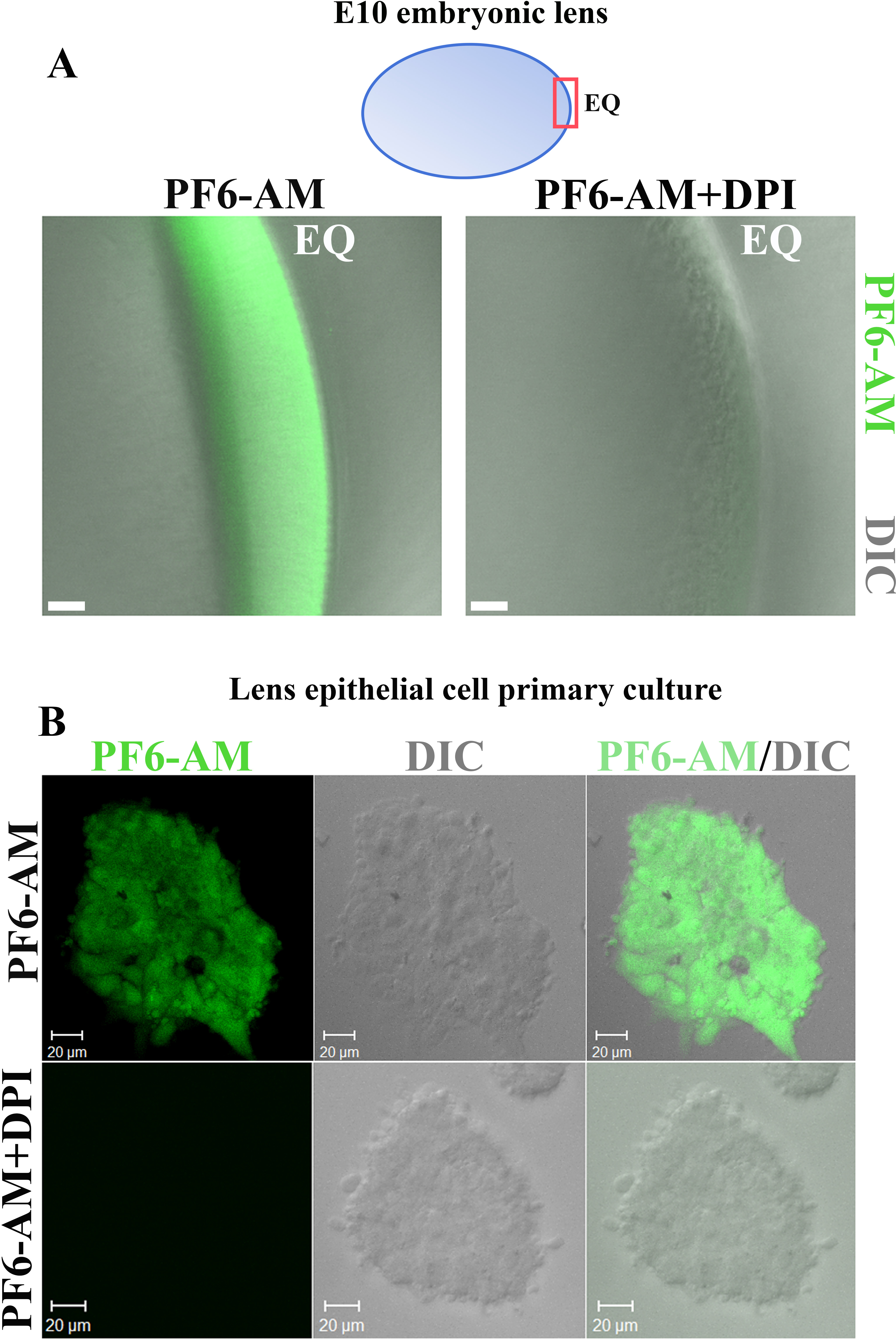
 Figure 2 of
Basu, Mol Vis 2014; 20:458-467.
Figure 2 of
Basu, Mol Vis 2014; 20:458-467.