Figure 6. Panel depicting the cellular distribution of pVHL in pterygia (A-C) and normal conjunctiva (D). Note the considerable staining heterogeneity in different pterygium samples (A-C). A: Weak cytoplasmic immunoreactivity in numerous epithelial cells of pterygium tissue. Arrows show basal epithelial cells with
increased staining compared to the other epithelial cells. B: Strong cytoplasmic immunoreactivity in basal and suprabasal cells of pterygium tissue. Arrows show pVHL positive stroma
vessels. C: The pVHL-expressing cells with strong immunoreactivity are distributed in all epithelial layers in this pterygium sample.
Arrows show pVHL-positive stroma vessels. Immunostaining is absent in sections incubated with control IgG (insert). D: Epithelial cells with weak cytoplasmic immunoreactivity are detected mainly in basal layer in normal conjunctival sample.
(Dako Envision Plus Detection System with hematoxylin counterstain, original magnification X400, bar 50 μm).
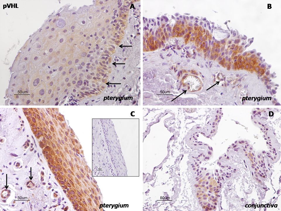
 Figure 6 of
Pagoulatos, Mol Vis 2014; 20:441-457.
Figure 6 of
Pagoulatos, Mol Vis 2014; 20:441-457.