Figure 5. Panel depicting the cellular distribution of Hsp70 in pterygia (A-D) and normal conjunctiva (E). A: Basal and suprabasal epithelial cells display moderate cytoplasmic Hsp70 immunoreactivity. B: Epithelium basal and suprabasal layers and stroma vessels (arrow) are Hsp70 positive in this pterygium sample. C: Epithelial cells distributed in the entire epithelium show Hsp70 immunoreactivity. Immunostaining is absent in sections
incubated with control IgG (insert). D: Basal cells exhibit more intense immunoreactivity compared to the other epithelial cells (arrows). E: Scattered basal epithelial cells with weak cytoplasmic immunoreactivity (arrows) are detected in normal conjunctival sample.
(Dako Envision Plus Detection System with hematoxylin counterstain, original magnification X400, bar 50 μm).
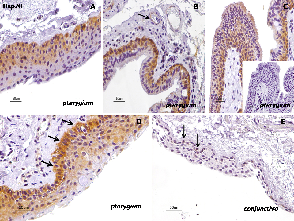
 Figure 5 of
Pagoulatos, Mol Vis 2014; 20:441-457.
Figure 5 of
Pagoulatos, Mol Vis 2014; 20:441-457.