Figure 10. Distribution of HIF-1α (green) and Hsp90 (red) as determined by double immunofluorescence staining in pterygia (Α-C) and normal conjunctiva samples (D-F). Nuclei are DAPI labeled in blue. A: Co-localization of HIF-1α nuclear expression with Hsp90 cytoplasmic expression in cells of the entire epithelium. B: Hsp90 is absent from focally distributed cells with cytoplasmic HIF-1α immunoreactivity. C: Cytoplasmic staining of HIF-1α is demonstrated in superficial layers of epithelium whereas Hsp90 staining is limited to
basal and suprabasal layers. D-F: Conjunctival basal epithelial cells demonstrate Hsp90 cytoplasmic immunoreactivity. E, F: A few HIF-1α positive cells with cytoplasmic staining are clearly detectable. Τhe dashed line indicates the pterygium basement
membrane (BM). The scale bar represents 50 μm.
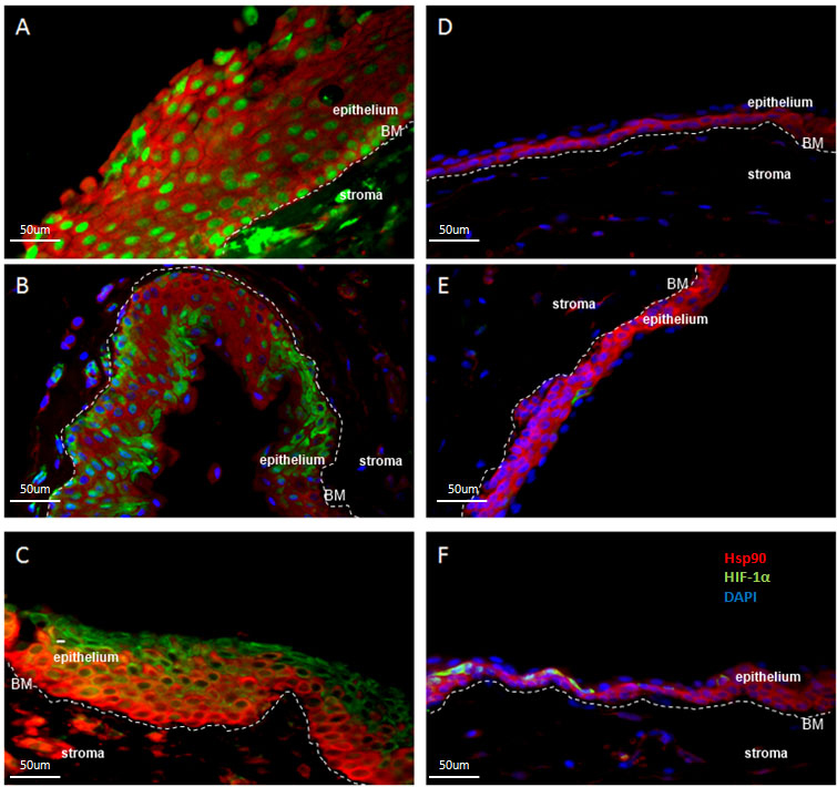
 Figure 10 of
Pagoulatos, Mol Vis 2014; 20:441-457.
Figure 10 of
Pagoulatos, Mol Vis 2014; 20:441-457.