Figure 2. Fundus changes in the proband with the heterozygous c.1619T>C (p.L540P) mutation in Family 2. A and B: RetCam examination of both of Patient 2’s eyes. The fundus of the left eye could not be viewed due to the opacity of the
lens. C and D: Ultrasound B- scan of Patient 2. These photos demonstrate leukocoria and severe retinal detachment of both eyes. The left
eye is more severely affected than the right eye. Posterior synechia is evident in the left eye. E: Fundus of Patient 2’s right eye at the last follow-up after lensectomy and vitrectomy. The retina was mostly attached. F-I: Fundus photos and fundus fluorescein angiography (FFA) of the asymptomatic mother with c.1619T>C in Family 2. F and G: Fundus photos show normal posterior fundi. H and I: FFA shows that the mother has peripheral nonperfusion areas, increased ramification, and shunts of the peripheral retinal
vessels in both eyes.
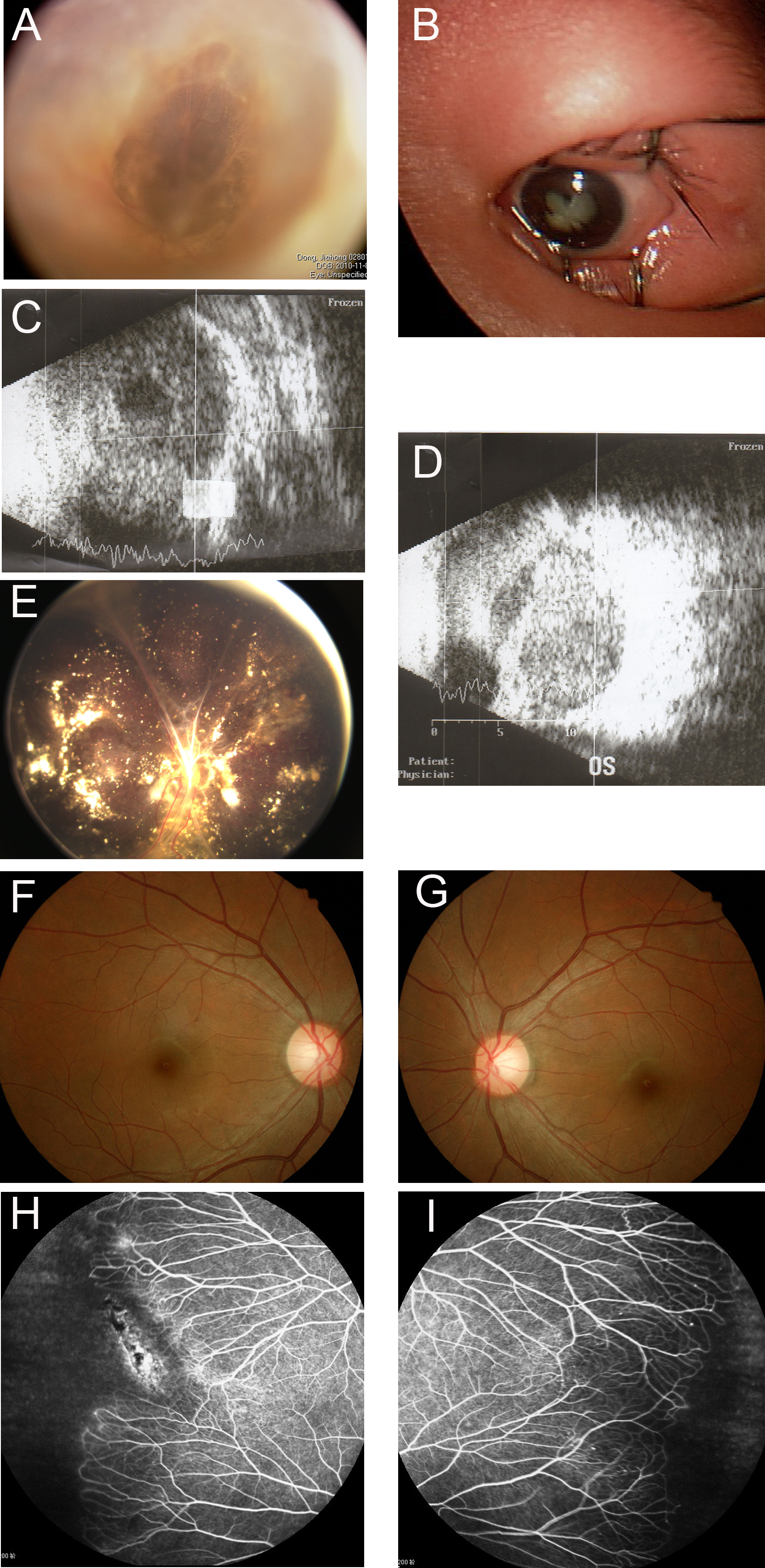
 Figure 2 of
Fei, Mol Vis 2014; 20:395-409.
Figure 2 of
Fei, Mol Vis 2014; 20:395-409.