Figure 1. Fundus changes in the proband with the heterozygous c.1264G>A (p.A422T) mutation in Family 1. A and B: The fundus of Patient 1 showed a retinal fold of the right eye and dragged disc of the left eye. C-F: Fundus photos of the asymptomatic father. C and D: Fundus photos of the asymptomatic father showed normal posterior fundi. E and F: Fundus fluorescein angiography (FFA) showed that the father had nonperfusion areas in both eyes.
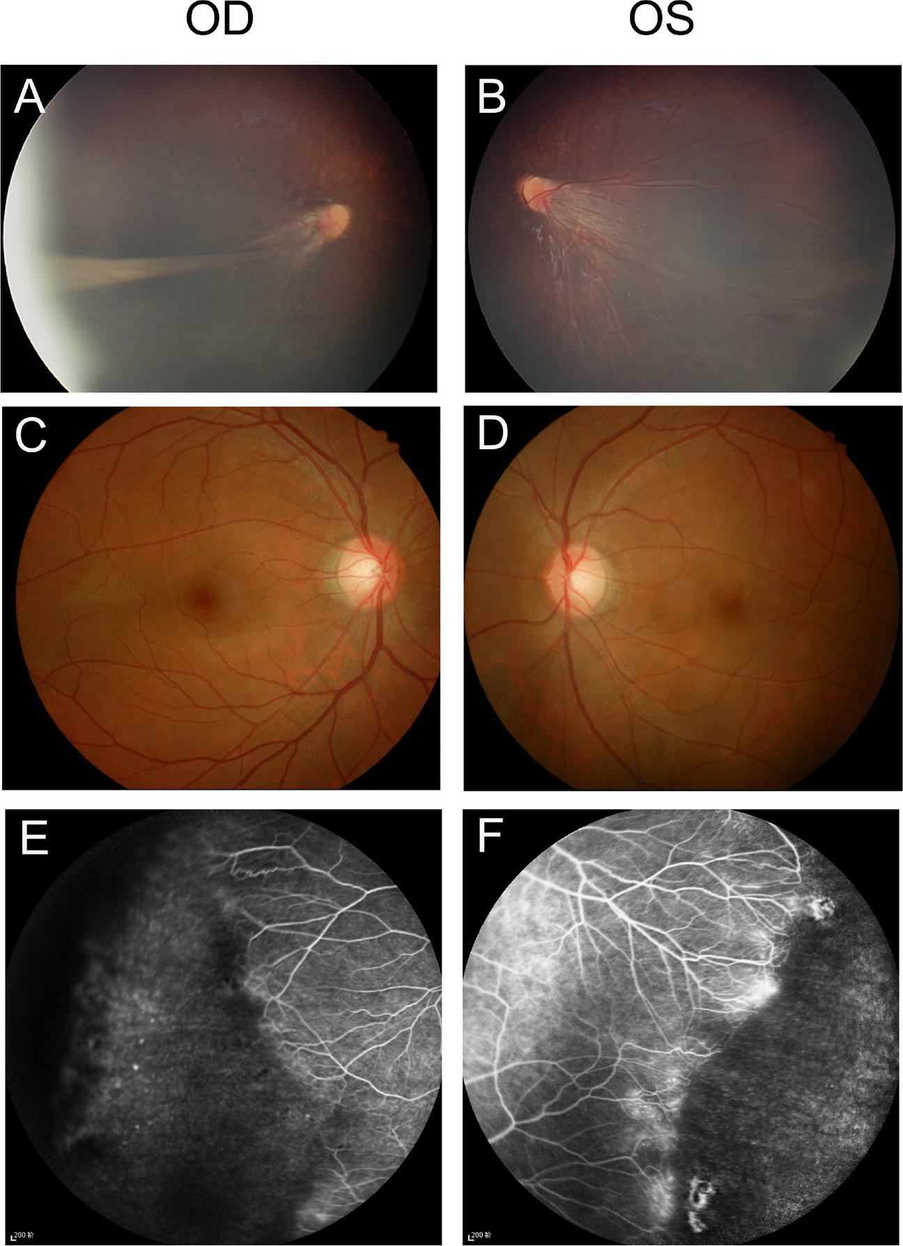
 Figure 1 of
Fei, Mol Vis 2014; 20:395-409.
Figure 1 of
Fei, Mol Vis 2014; 20:395-409.