Figure 3. Histology and immunology of native and reconstructed corneas. Native (A, D, G, J and M) and reconstructed corneas were sectioned for microscopic observation. Corneal constructs contained human endothelium directly
seeded under the collagen gel (B, E, H, K and N) or bovine endothelium growing on a synthetic membrane (C, F, I, L and O). As shown on the paraffin sections, stained with Masson’s trichrome (A–C), cells are stained pink, and the collagen tissue is blue. Immunofluorescent images of cryosections (D–O) have been exposed to the antibodies against the following antigens: α-catenin (D–F), β-catenin (G–I), E-cadherin (J–L), and N-cadherin (M–O). Cell nuclei were stained blue with Hoechst dye (D–R). Scale bar = 50 µm.
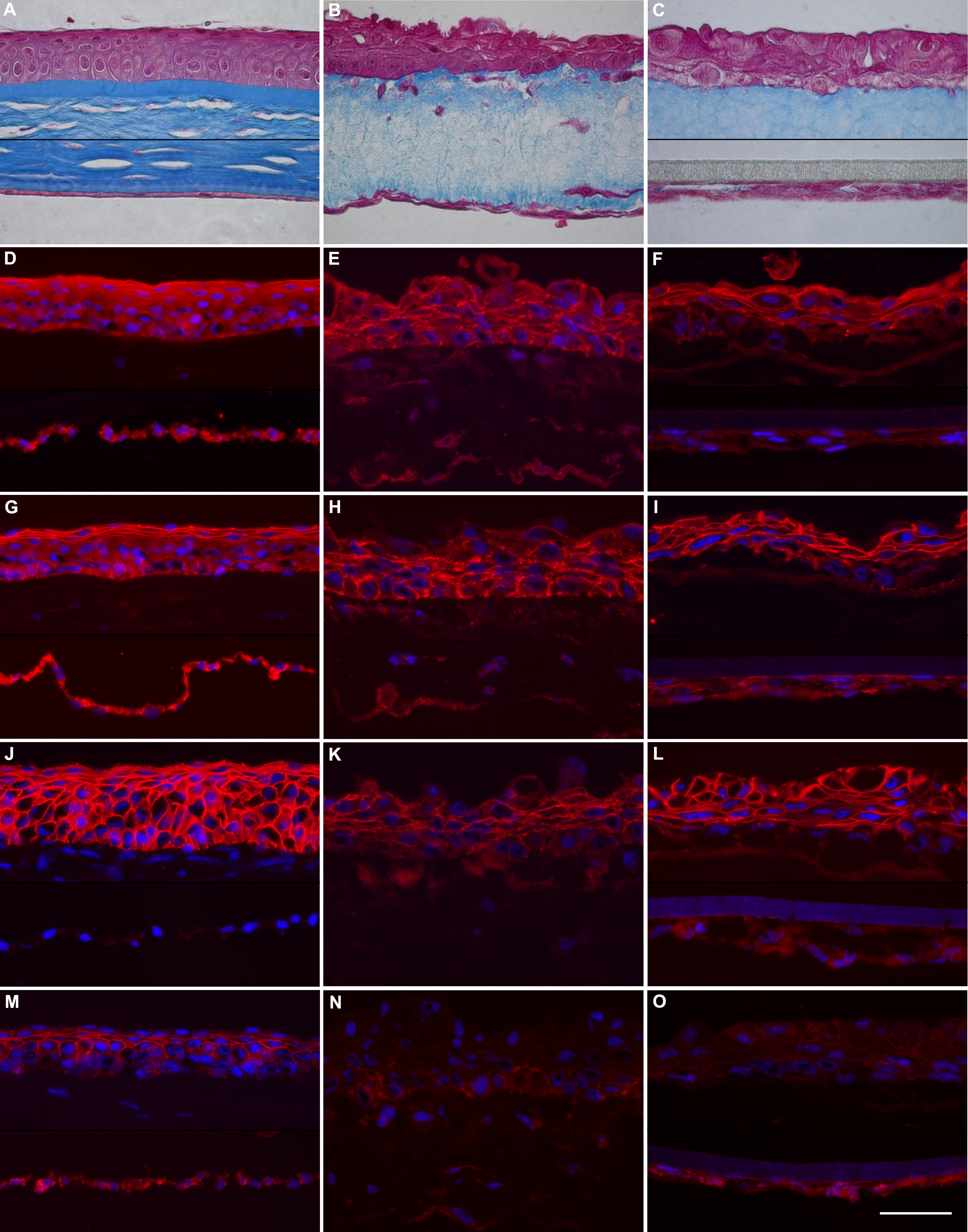
 Figure 3 of
Giasson, Mol Vis 2014; 20:386-394.
Figure 3 of
Giasson, Mol Vis 2014; 20:386-394.