Figure 1. Schematic diagram of the preparation of corneal equivalent made with collagen. Cells were harvested from human corneas and
separately grown in culture dishes. Just before confluence, cells were trypsinized to produce a suspension. Cells were added
over the collagen construct cast in a Petri dish (model 1). Alternatively, endothelial cells obtained from bovine corneas
were seeded over a culture insert to produce a collagen corneal construct made of human epithelium and keratocytes (model
2).
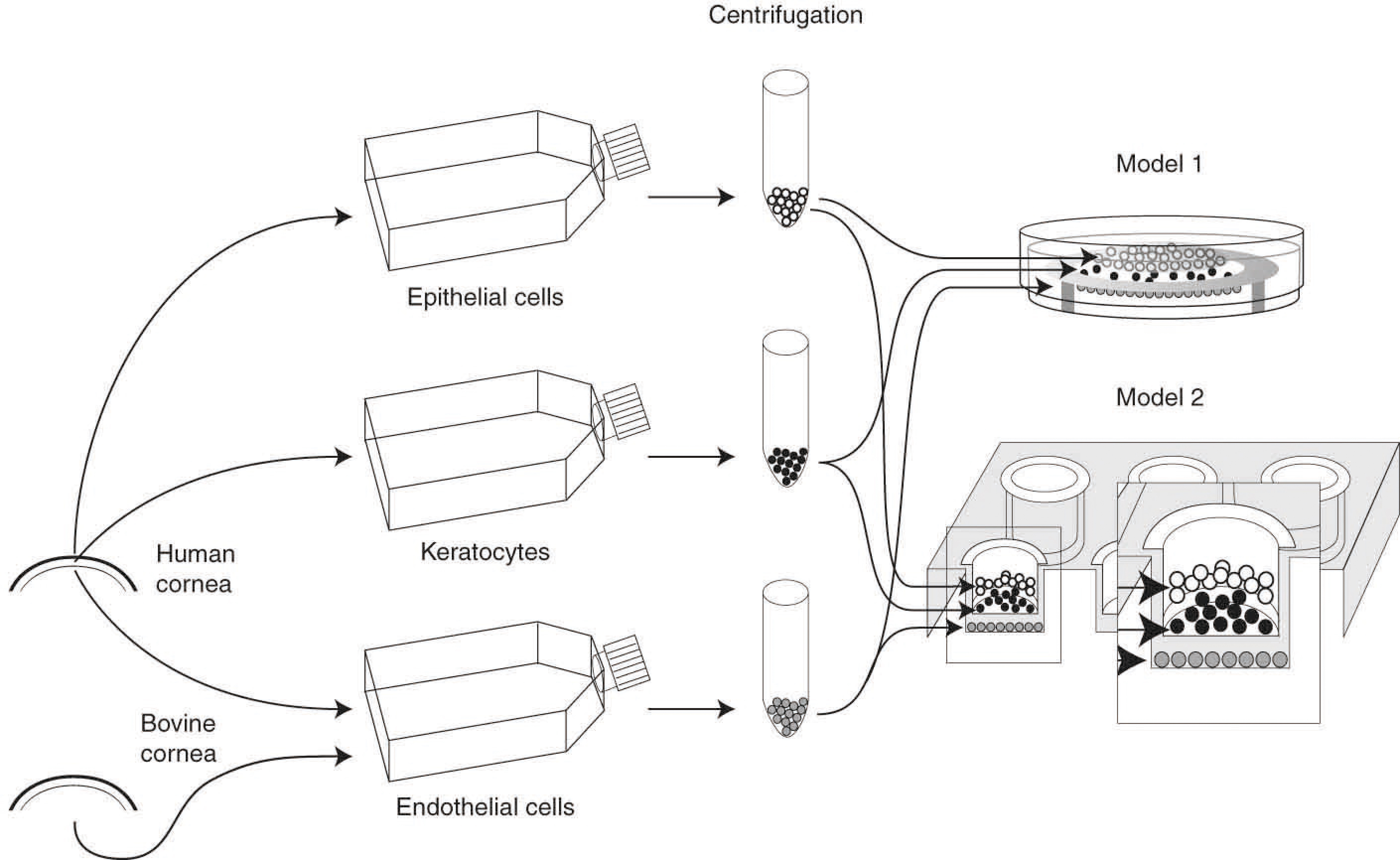
 Figure 1 of
Giasson, Mol Vis 2014; 20:386-394.
Figure 1 of
Giasson, Mol Vis 2014; 20:386-394.