Figure 4. Magnetic resonance imaging. A: T1-weighted axial magnetic resonance imaging (MRI) shows diffuse thinning of the extraocular muscles. B: Sagittal diffusion tension imaging (DTI) demonstrates a normal corpus callosum configuration. C: T2-weighted coronal MRI illustrates a normal optic chiasm.
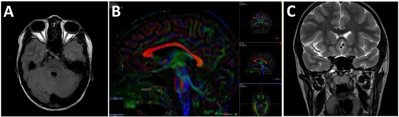
 Figure 4 of
Ali, Mol Vis 2014; 20:368-375.
Figure 4 of
Ali, Mol Vis 2014; 20:368-375.