Figure 4. Fundus photography and perimetry of the two children. Retinal pigment epithelium mid-periphery pigment changes are already
visible. Perimetry shows concentric reduction in the visual field in patient III:1 (A, B) and patient III:2 (C, D). RE, right eye; LE, left eye.
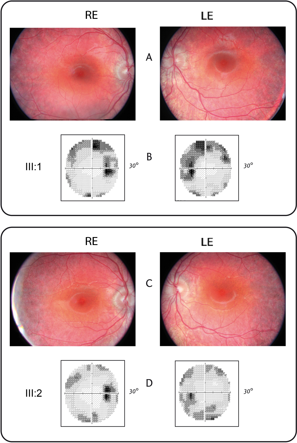
 Figure 4 of
Contestabile, Mol Vis 2014; 20:325-333.
Figure 4 of
Contestabile, Mol Vis 2014; 20:325-333.