Figure 2. Optical coherence tomography, visual field, and fluorescein angiography of the proband and his brother. Widespread chorioretinal
atrophy with relative sparing of the macular region and visual field constriction are found. Note the greater severity of
the anatomic and functional findings of the proband II:4 (A, B, C) compared to his older brother II:3 (D, E, F). RE,right eye; LE, left eye.
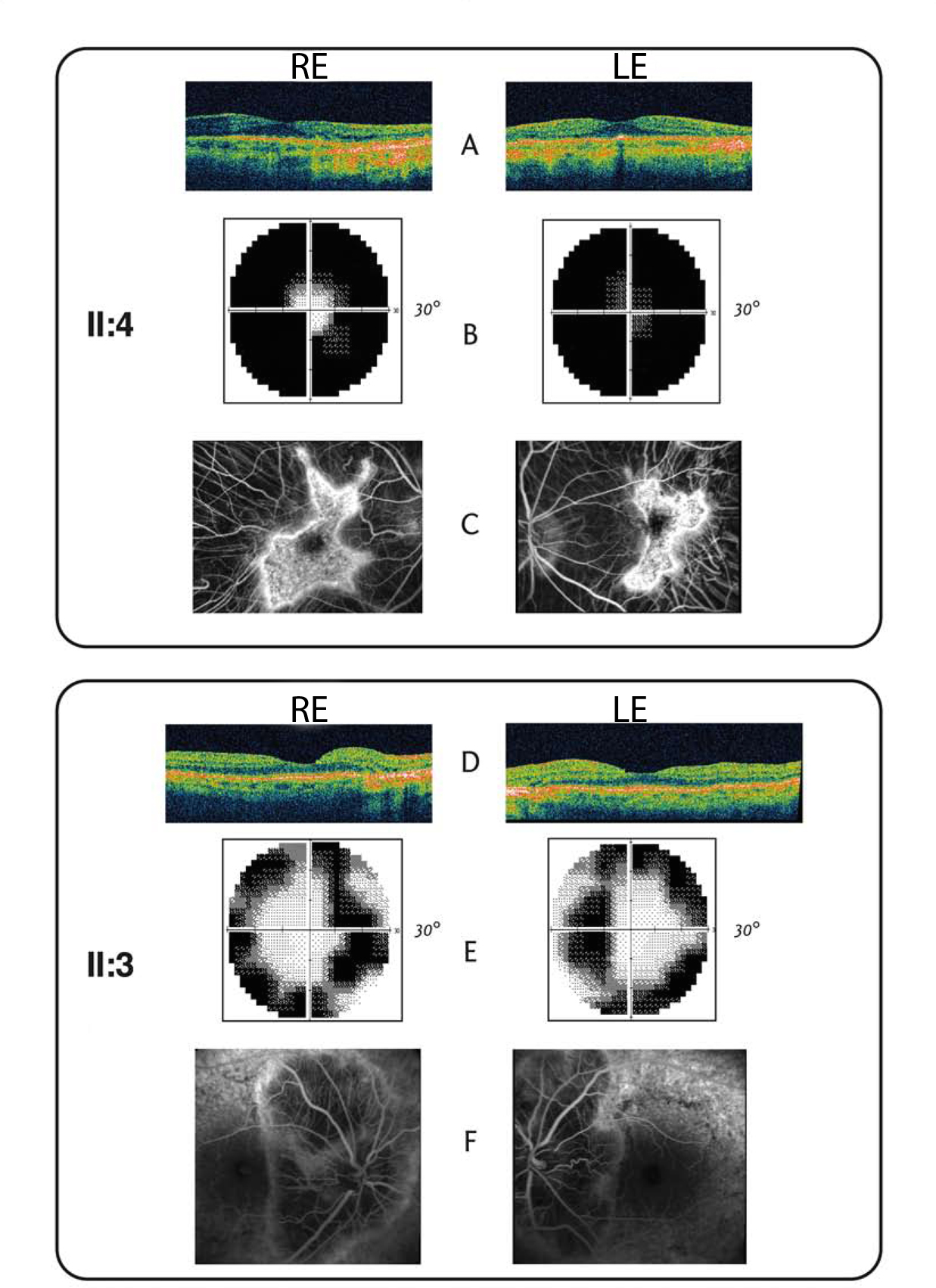
 Figure 2 of
Contestabile, Mol Vis 2014; 20:325-333.
Figure 2 of
Contestabile, Mol Vis 2014; 20:325-333.