Figure 2. Partial colocalization of A2E and LysoTracker fluorescence in A2E-fed ARPE-19 cells (60× magnification). A: A2E autofluorescence; B: 4',6-diamidino-2-phenylindole dihydrochloride (DAPI); C: LysoTracker Red DND 99; and D: merged. Confocal microscopy was performed on an Olympus FV1000 confocal microscope with detection in DAPI, green (fluorescein
isothiocyanate, FITC), and red (rhodamine) channels as described. Results shown are representative of two separate experiments.
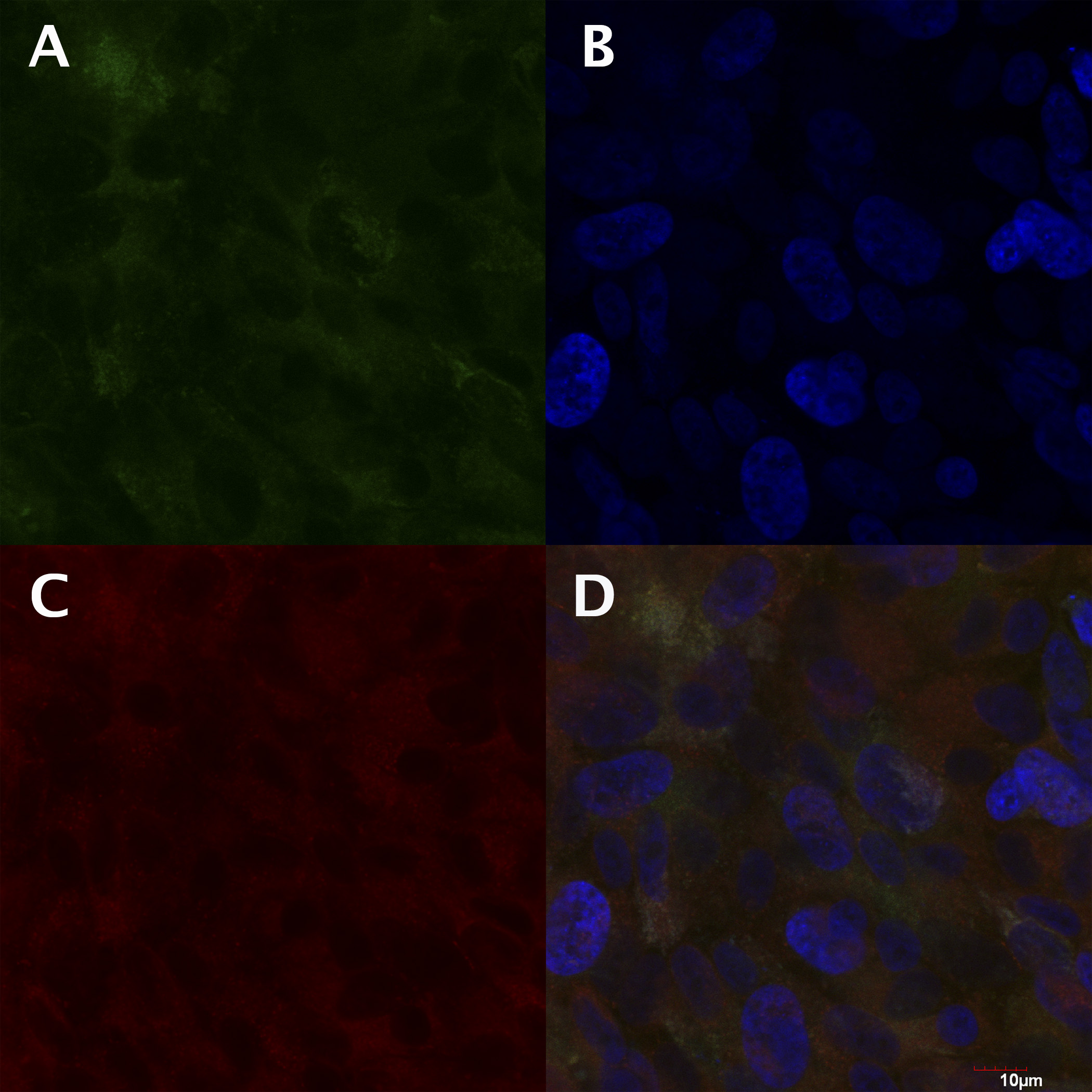
 Figure 2 of
Poliakov, Mol Vis 2014; 20:285-300.
Figure 2 of
Poliakov, Mol Vis 2014; 20:285-300.