Figure 2. Transcriptional regulation by PAX6 and PAX6(5a) on motif 1–1, 1–2, and 1–3 luciferase reporters. A: Distribution of the nucleotides in the Pax6-binding motifs 1–1, 1–2, and 1–3. The DNA sequences for gene synthesis are aligned
with the corresponding motifs. The binding sites and flanking sequences are in upper and lower case, respectively. Evaluation
of PAX6 (black bars) and PAX6(5a) (gray bars) series with 1–1, 1–2, and 1–3 luciferase reporters in cotransfected P19 cells
is shown in panel (B), (C), and (D) respectively. The sample size n=6 from two independent triplicates. The error bars represent the standard deviation. The
significant fold-changes are indicated by the asterisks. The range of the p values are: *** <0.001, ** 0.001~0.01, * 0.01~0.05.
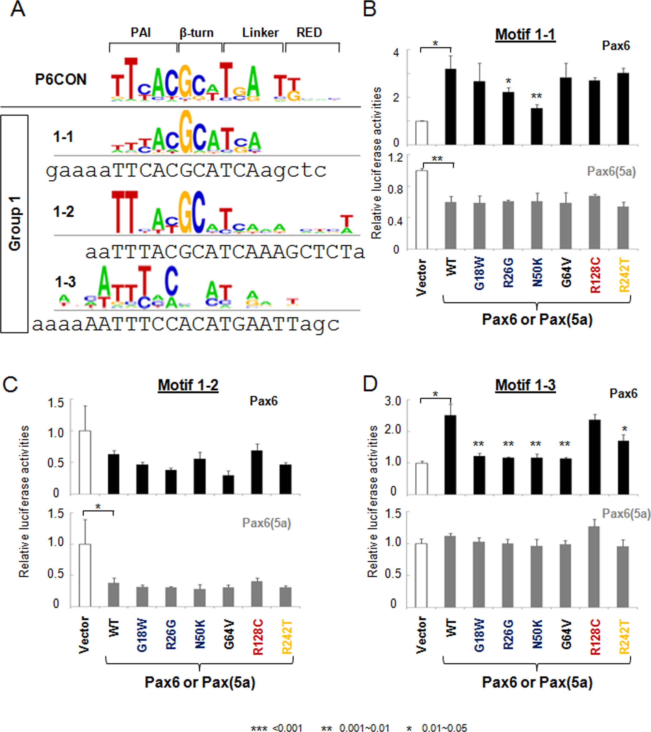
 Figure 2 of
Xie, Mol Vis 2014; 20:270-282.
Figure 2 of
Xie, Mol Vis 2014; 20:270-282.