Figure 8. Mechanisms of myricetin-induced retinal pigment epithelial cell necrosis. The rate of DNA fragmentation in the cultured media
was determined after stimulation of the cells with myricetin for 6 h (A) and 24 h (B), respectively. The following agents were tested in the absence (control) and presence of myricetin (200 µM): the caspase-3
and -8 specific inhibitors DEVD (100 μM) and IETD (100 µM), the poly(ADP-ribose) polymerase-1 (PARP-1) inhibitor 3,4-dihydro-5-[4-(1-piperidinyl)butoxy]-1(2H)-isoquinoline
(DPQ; 30 µM), the inhibitor of the nicotinamide adenine dinucleotide phosphate-oxidase (NADPH) oxidase pathway and the uncoupling
protein-2/mitochondrial pathway, perindopril (4 µM), the inhibitor of the mitochondrial permeability transition cyclosporin
A (CsA; 1 µM), the mitochondrial KATP channel opener pinacidil (10 µM), the calpain inhibitor PD150606 (100 µM), the reducing agent dithiothreitol (DTT; 3 mM),
the inhibitor of phospholipase A2 4-bromophenacyl bromide (Bromo; 300 µM), the cyclooxygenase inhibitor indomethacin (Indo; 10 µM), the mitogen-activated protein
kinase (MEK) inhibitor, PD98059 (20 µM), and the c-Jun NH2-terminal kinase (JNK) inhibitor, SP600125 (10 µM). Data are means±standard error of the mean (SEM) of three to six independent
experiments using cells from different donors, and are expressed in percent of untreated control (100%). Significant difference
vs. untreated control: *p<0.05. Significant difference vs. myricetin control: ●p<0.05.
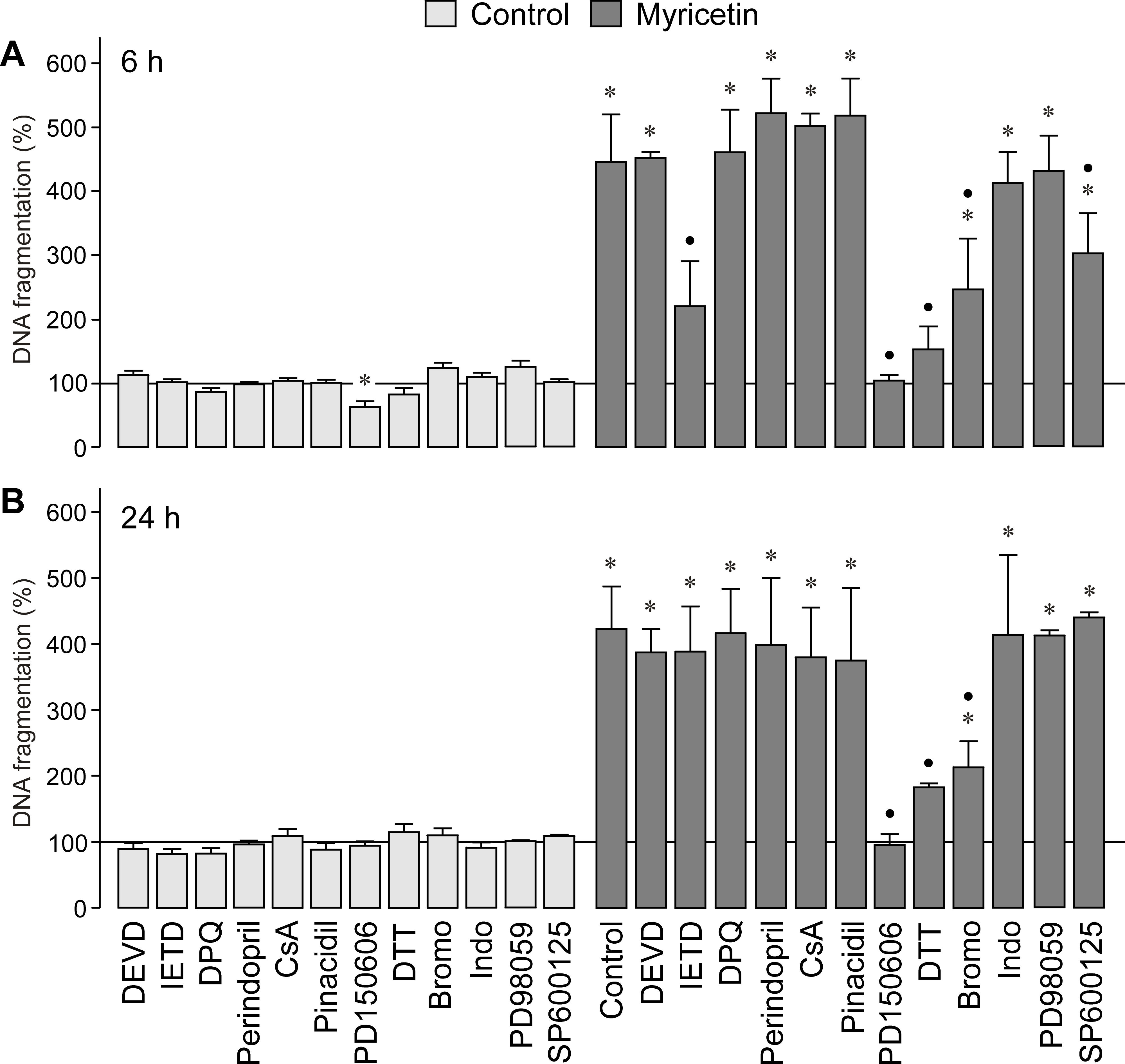
 Figure 8 of
Chen, Mol Vis 2014; 20:242-258.
Figure 8 of
Chen, Mol Vis 2014; 20:242-258.