Figure 7. Dose-dependent effects of several vegetable polyphenols on the rate of DNA fragmentation of retinal pigment epithelial (RPE)
cells. (A) epigallocatechin-3-gallate, (B) luteolin, (C) apigenin, (D) myricetin, (E) quercetin, and (F) cyanidin. The rate of DNA fragmentation was determined in the cell lysates (to determine the level of cellular apoptosis)
and culture supernatants (to determine the level of cellular necrosis) after 6 h (left side) and 24 h of cell culturing (right
side). Triton X-100 (1%) was used as positive control. The vehicle controls were made with acetone (Ac; 0.2%), dimethyl sulfoxide
(DMSO; 0.2%), and methanol (Me; 0.4%), respectively. The concentrations of the test substances (in µM) are given in the bars.
Data are means±standard error of the mean (SEM) of three to six independent experiments using cells from different donors,
and are expressed in percent of untreated control (100%). Significant difference vs. untreated control: *p<0.05.
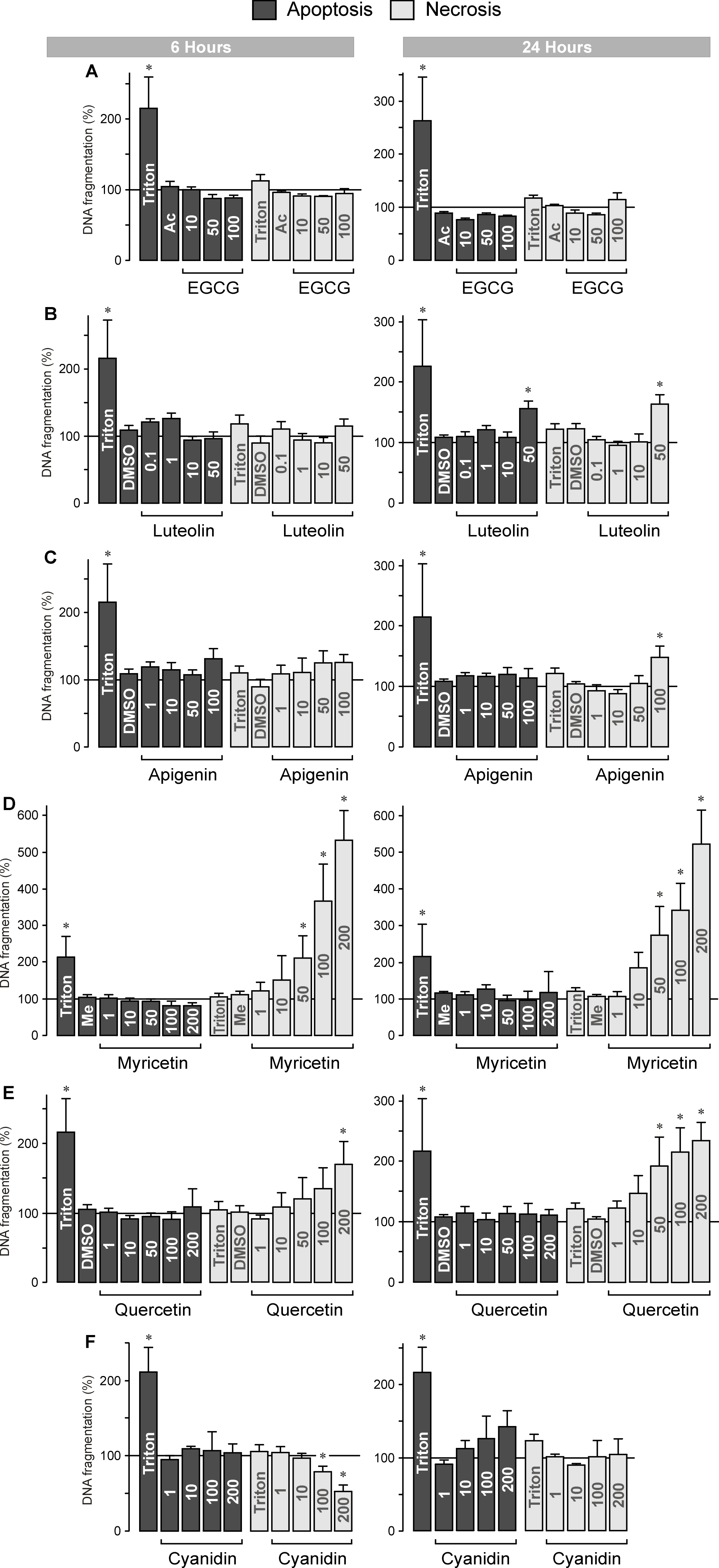
 Figure 7 of
Chen, Mol Vis 2014; 20:242-258.
Figure 7 of
Chen, Mol Vis 2014; 20:242-258.