Figure 7. Changes in the expression of cell cycle progressing cyclins and cyclin-dependent kinases in the presence of hepatocyte growth
factor (HGF) and keratinocyte growth factor (KGF). Rabbit corneal primary epithelial (RCPE) cell growth cultures at 80%–90%
confluence were starved overnight with Dulbecco’s Modified Eagle Medium with Ham’s F-12 (DMEM/F-12)/0.25% fetal calf serum
(FCS) and then treated with 20 ng/ml HGF or KGF for 10 h (A) or 24 h (B). Levels of cyclins A, D, and E, and cyclin-dependent kinase 2 (CDK2) and CDK4 in cellular extracts were determined with
western immunoblotting by using polyclonal antibodies specific for each protein. Data presented represent results obtained
from two to three similar experiments. Quantification of cell cycle proteins is shown in bar diagrams (C). Data shown are mean±standard deviation (SD). *p<0.05, control versus HGF or KGF at 10 or 24 h.
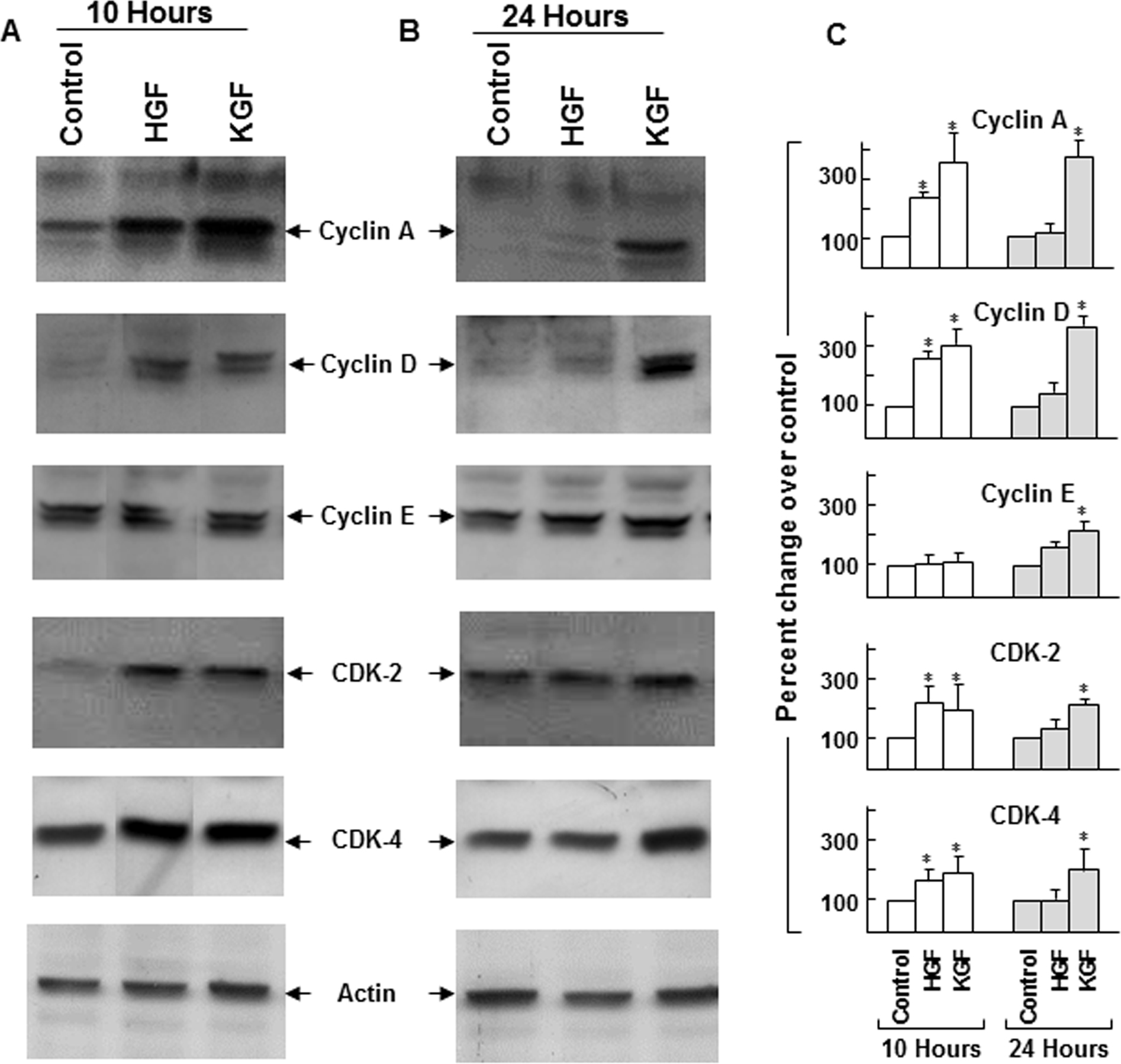
 Figure 7 of
Chandrasekher, Mol Vis 2014; 20:24-37.
Figure 7 of
Chandrasekher, Mol Vis 2014; 20:24-37.