Figure 3. Changes in the levels of p27kip and p21cip during apoptosis. A, B: Apoptosis was induced in rabbit corneal primary epithelial (RCPE) cell cultures with staurosporine (STS) as described in
the Methods section. E: Subconfluent (80%–90%) RCPE cultures were starved overnight with Dulbecco’s Modified Eagle Medium with Ham’s F-12 (DMEM/F-12)/0.25%
fetal calf serum (FCS) and then treated with 20 ng/ml HGF or KGF for 10 or 24 h. Levels of p27kip and p21cip in cellular extracts at different time points were determined with western immunoblotting using specific antibodies. Quantification
of p27kip and p21cip levels in apoptosis cultures (C–D) and HGF- and KGF-treated cultures (F) was performed with densitometry. Data are representative of results obtained from three similar experiments. Data shown
are mean±standard deviation (SD). *p<0.05, STS versus corresponding control at 10 or 24 h, #p<0.05, control versus HGF or
KGF at 10 or 24 h.
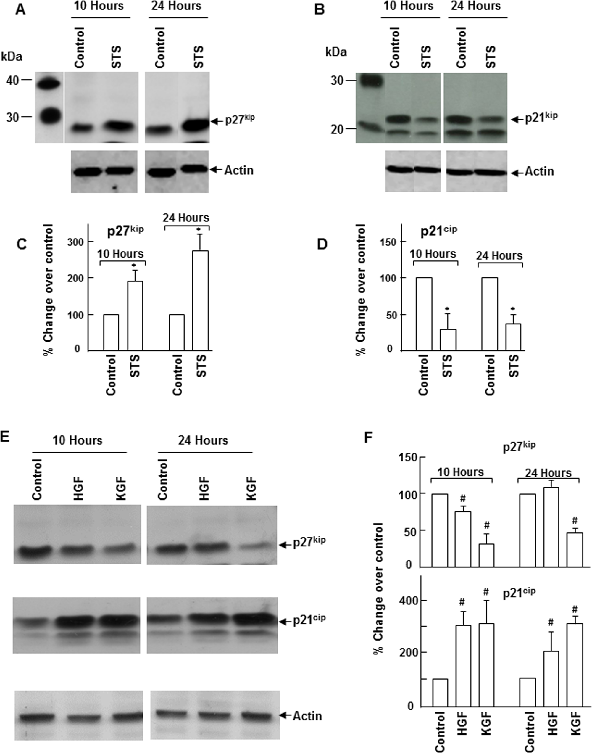
 Figure 3 of
Chandrasekher, Mol Vis 2014; 20:24-37.
Figure 3 of
Chandrasekher, Mol Vis 2014; 20:24-37.