Figure 1. Hepatocyte growth factor (HGF)– and keratinocyte growth factor (KGF)–mediated cell survival during staurosporine (STS)-induced
apoptosis. Rabbit corneal primary epithelial (RCPE) cell cultures in Dulbecco’s Modified Eagle Medium with Ham’s F-12 (DMEM/F-12)/0.25%
fetal calf serum (FCS) were pretreated with 20 ng/ml HGF or KGF for 30 min before STS was added. Cultures were further incubated
in the presence of STS (10 ng/ml) for 2 h. Then the medium was removed, and fresh medium containing HGF or KGF but no STS
was added, and incubation continued for an additional 2 to 22 h. The cultures were stained with Hoechst fluorescent nuclear
staining reagent to visualize cells containing apoptotic nuclei as described in the Methods section, and images were captured
(A). The number of apoptotic cells in each experimental condition was quantified and expressed as the percentage of total cells
(apoptotic + normal; B). Data shown are mean±standard deviation (SD). *p<0.05, STS alone versus various treatments after 4 or 10 or 24 h.
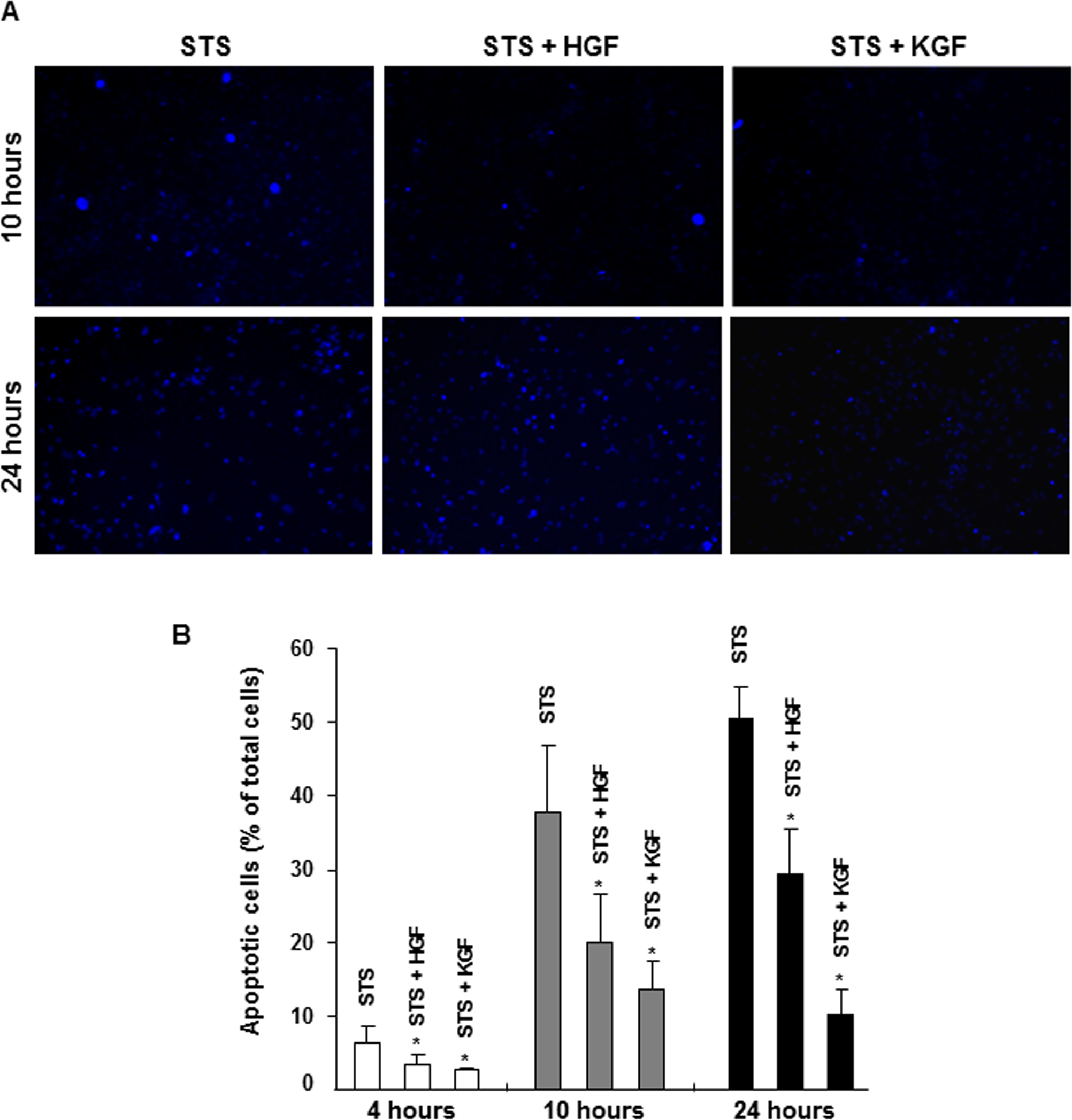
 Figure 1 of
Chandrasekher, Mol Vis 2014; 20:24-37.
Figure 1 of
Chandrasekher, Mol Vis 2014; 20:24-37.