Figure 6. Histological studies reveal a range of disease severity in the eyes of chimeric mice. A-H: Representative photomicrographs of hematoxylin and eosin stained sections of chimeric (A, C, E) and Arf −/− (G) eyes show a retrolental mass (RLM; shown at higher magnification in B, D, F, H) with dense accumulation of pigmented cells. I: Tabulation of whether a RLM is present in the midline sections of individual eyes. J: Mean size of the RLM is larger in Arf −/− mice than in chimeric mice (p = 0.031). Error bars depict standard deviation.
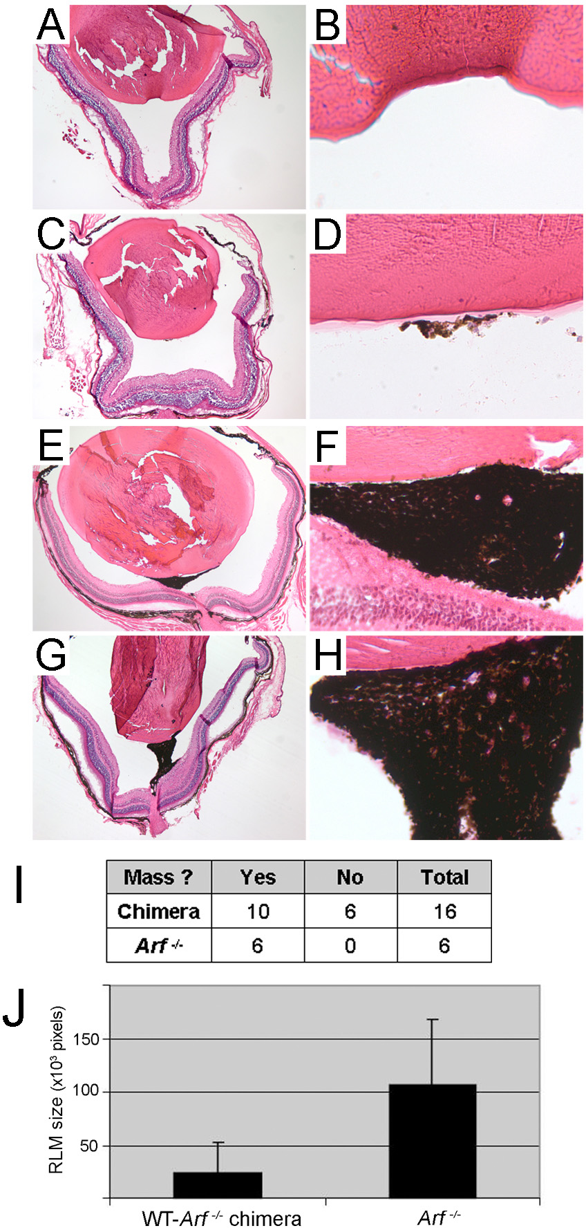
 Figure 6 of
Mary-Sinclair, Mol Vis 2014; 20:215-230.
Figure 6 of
Mary-Sinclair, Mol Vis 2014; 20:215-230.