Figure 5. Ophthalmoscopy evaluation of chimeric mice shows a range of disease severity. A: Tabulation showing the number of eyes with low, moderate, and high degree of disease (L, M, H, respectively) based on slit-lamp
and fundoscopic evaluations. Note that two eyes included in the Low category were felt to be normal by visual inspection.
B: Representative photographs of eyes used to generate data in (A). The black arrow indicates a hyaloid vessel. White arrows denote focal lens opacity.
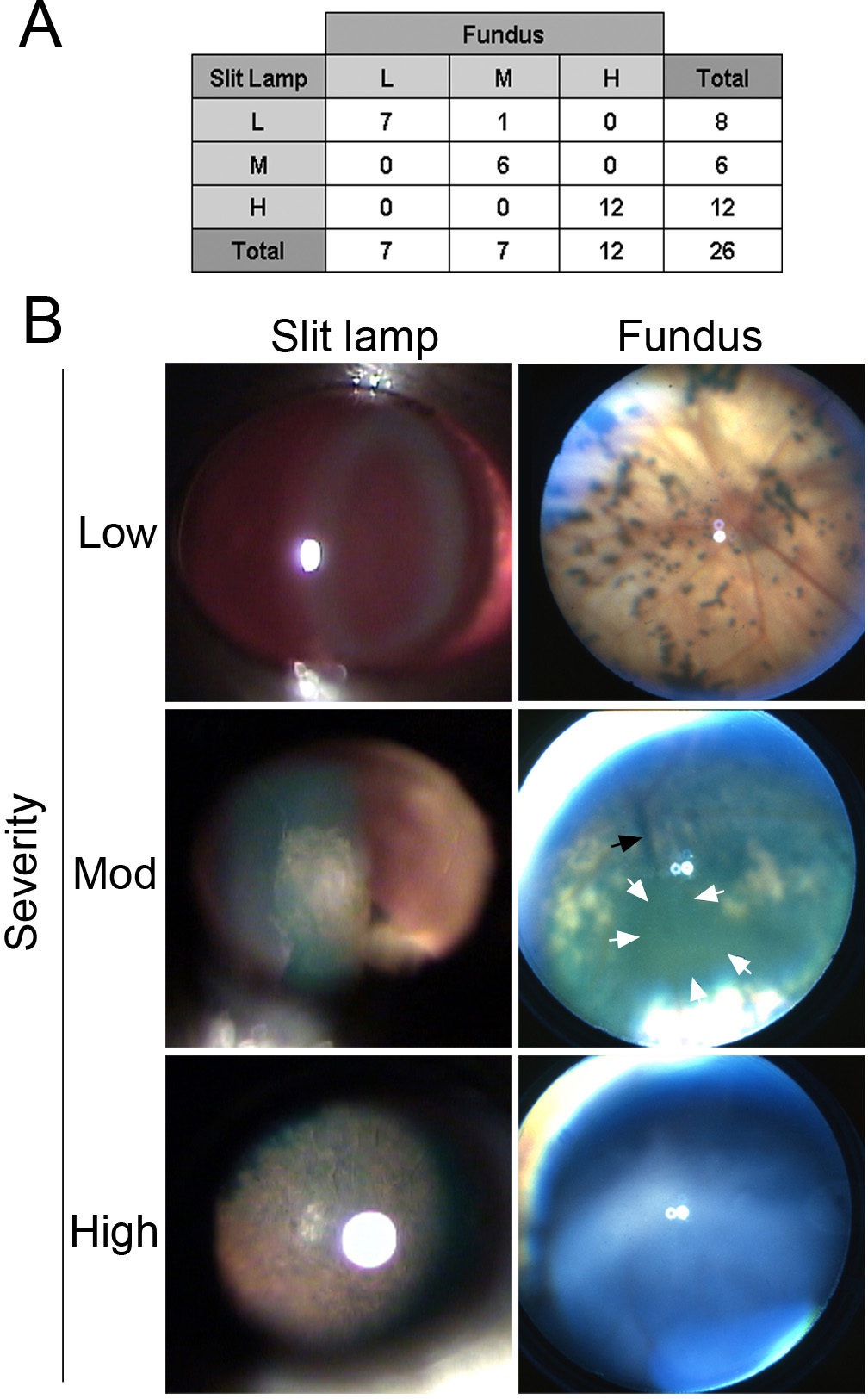
 Figure 5 of
Mary-Sinclair, Mol Vis 2014; 20:215-230.
Figure 5 of
Mary-Sinclair, Mol Vis 2014; 20:215-230.