Figure 2. Eyes from chimeric mice are composed of varying degrees of wild type and Arf −/− cells. A-H: Representative photomicrographs of fluorescence (A-F) or light microscopic images of hematoxylin and eosin stained sections (G and H). Letters (H, M, K, A) to the side of images indicate the animal ID. Green fluorescence (A, C, E) denotes wild type lineage; 4',6-diamidino-2-phenylindole dihydrochloride (DAPI) staining (B, D, F) shows all nuclei in sections. Arrows and arrowheads show the corneal epithelium and endothelium, respectively (A-F) and wild type (non-pigmented) and Arf −/− (pigmented) RPE, respectively (G and H). Insets (A-F) show higher magnification. I: Absolute quantification of the relative contribution from the Arf-/- lineage in the cornea and the RPE, represented as percentage of total. Letters and letter primes distinguish individual eyes
from the same chimeric animal.
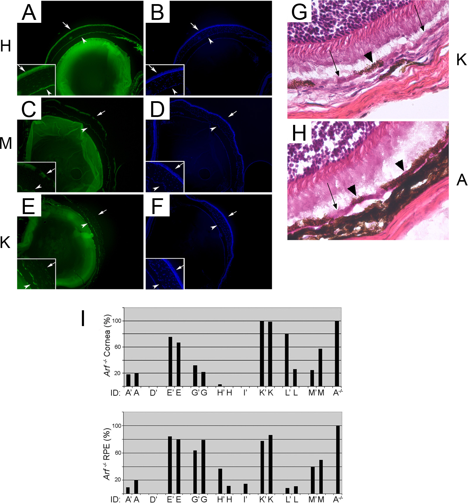
 Figure 2 of
Mary-Sinclair, Mol Vis 2014; 20:215-230.
Figure 2 of
Mary-Sinclair, Mol Vis 2014; 20:215-230.