Figure 2 of
Meng, Mol Vis 2014; 20:1806-1814.
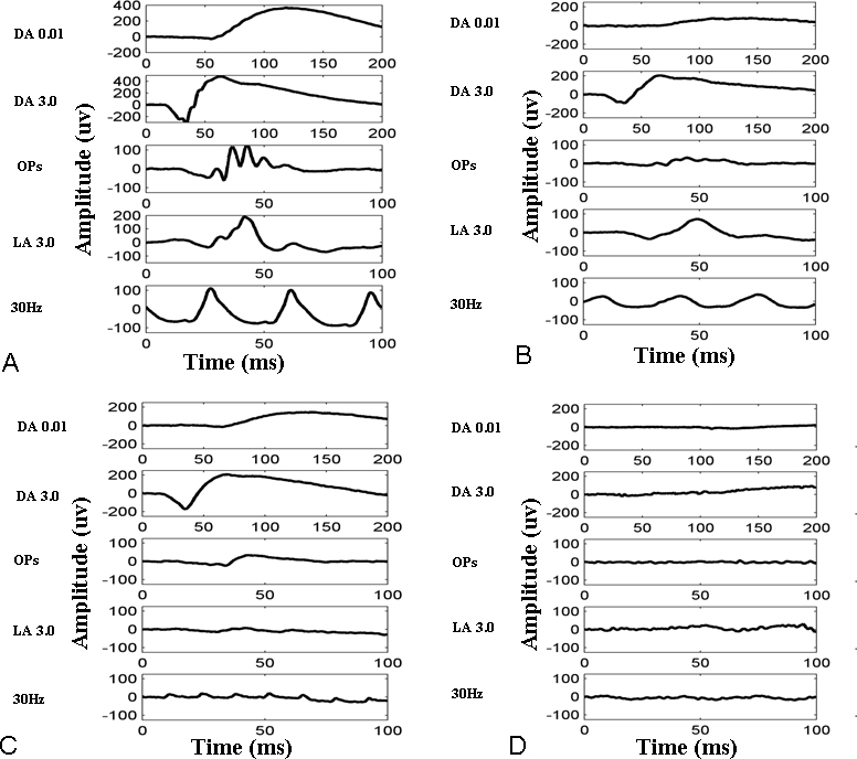
Figure 2.
Full-field ERG of normal subjects and BCD patients.
A
: Normal electroretinogram (ERG) from age-matched subject.
B
: Cone dominant ERG.
C
: Rod dominant ERG.
D
: Non-recordable ERG from patient 33, patient 60, and patient 86.
 Figure 2 of
Meng, Mol Vis 2014; 20:1806-1814.
Figure 2 of
Meng, Mol Vis 2014; 20:1806-1814.