Figure 1 of
Zobor, Mol Vis 2014; 20:178-182.
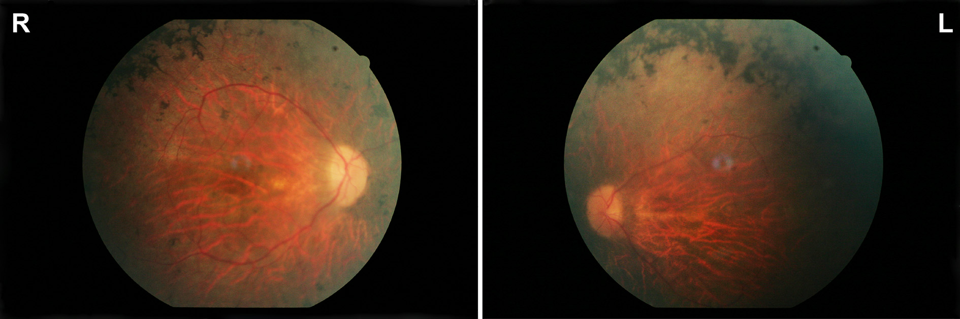
Figure 1.
Morphological findings. Fundus pictures of the right (R) and left eye (L) of the index patient showing pallor optic discs, attenuated vessels, and characteristic pigment changes in the mid-periphery.
 Figure 1 of
Zobor, Mol Vis 2014; 20:178-182.
Figure 1 of
Zobor, Mol Vis 2014; 20:178-182.