Figure 9. Representative LSCM images of severely damaged RCS dystrophic lenses at 7–8 weeks postnatal. In a subset of lenses (~25%),
many posterior fiber ends adopted a stellate shape. A–D: The same field of view showing F-actin (A), cadherin (B), integrin (C), and the merged image of the triple-labeled fiber ends (D). Orientation is en face. F actin was localized to the stellate arms of the fiber ends (A, D, arrows); however, integrin had a diffuse distribution with more pronounced labeling in the stellate arms (C). Cadherin was localized to irregularly shaped plaques between stellate projections (B, asterisks). E–F: Anterior (E) and posterior (F) polar sections doubled labeled with phalloidin-FITC and DAPI demonstrate the distribution of F-actin and nuclei. Nuclei
were present only in anterior sections (E, stars); no DAPI-positive fluorescence was present in posterior sections. F-actin was localized to stellate projections (F, arrows). This confirms that the cadherin-positive plaques do not represent nuclei. Panels A–F are at the same magnification.
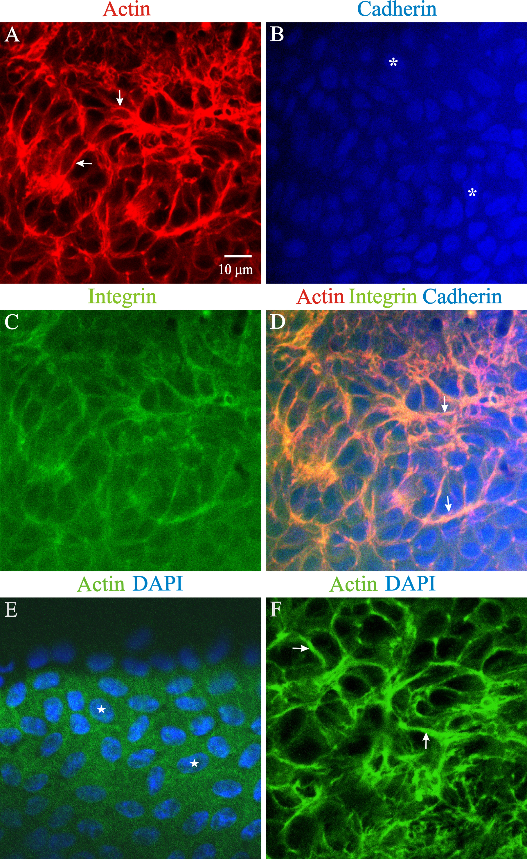
 Figure 9 of
Joy, Mol Vis 2014; 20:1777-1795.
Figure 9 of
Joy, Mol Vis 2014; 20:1777-1795.