Figure 8. Diagram summarizing the distribution of vinculin (shown in gold; A-D) and β integrin (shown in green; E-H) in the BMC of migrating fiber ends in RCS/Lav (dystrophic) rats. A, E: At 2 weeks old; B, F: at 4 weeks old; C, G: at 6 weeks old; D, H: at 8 weeks old. At 2 weeks postnatal, vinculin was distributed as scattered punctuate spots; however, this pattern was altered
by 4 weeks postnatal. Specifically, many fiber ends displayed diffuse strata of vinculin within the BMC, and this pattern
persisted through 6 weeks postnatal. In the majority of 8-week-old dystrophic lenses, immunofluorescence for vinculin was
arranged as small punctate foci around the border of basal fiber ends. At 2–4 weeks postnatal, β integrin was distributed
as discrete spots, often coincident with the BMC borders. However, in lenses from 6- to 8-week-old dystrophic rats, faint
immunofluorescence for β integrin was present in the cytoplasm and to a greater degree around the periphery of basal fiber
ends.
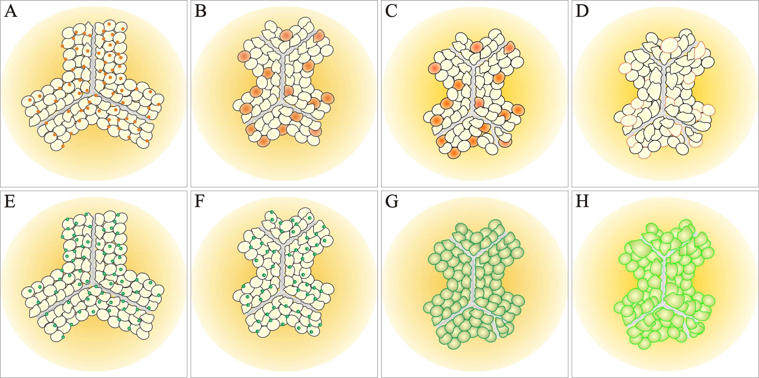
 Figure 8 of
Joy, Mol Vis 2014; 20:1777-1795.
Figure 8 of
Joy, Mol Vis 2014; 20:1777-1795.