Figure 1. Diagrammatic representation of the orientation of vibratome sections, location of intact posterior fiber ends, and region
where imaging of the BMC was performed. A: Posterior polar sections were obtained with a vibrating knife microtome, yielding serial sections of increasing diameter.
B: An enlarged edge-on view of the first two sections demonstrates the location of intact migrating basal fiber ends. In the
initial section, intact basal fiber ends were present beneath the entire curved surface of the enveloping capsule, whereas
all subsequent sections only had intact fiber ends beneath the beveled edge covered by the capsule. Sections are not to scale.
C: Enlarged en face view of the posterior surface of the lens shows basal fiber ends beneath the capsule in the first section
(blue dashed line) and in subsequent sections (green and red dashed lines). The regions of fiber-end migration where the BMC
architecture was imaged are encompassed within the outermost red dashed line and comprise the distal portion of the lateral-posterior
region, the perisutural region, and the sutural region.
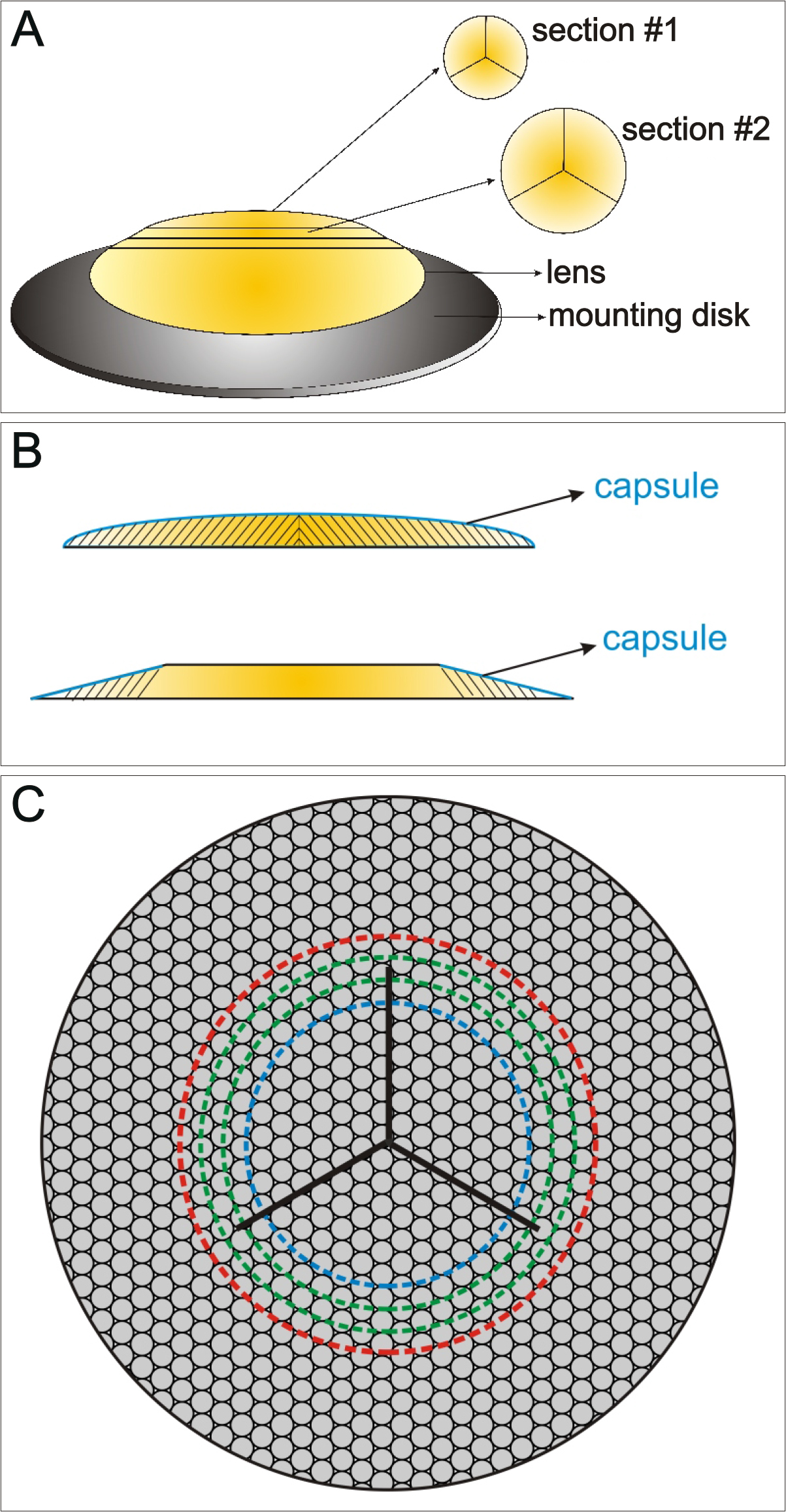
 Figure 1 of
Joy, Mol Vis 2014; 20:1777-1795.
Figure 1 of
Joy, Mol Vis 2014; 20:1777-1795.