Figure 1. The expression of RAGE in human pterygium and normal conjunctiva tissues. A, B: Immunohistochemistry (IHC) staining of RAGE in conjunctiva and pterygium specimens. RAGE is expressed in endothelial cells
lining the lumen of blood vessels (black arrows). In addition, other cell types also exhibit RAGE expression, notably epithelial
cells (black arrow heads), fibroblasts (red arrows), and inflammatory cells (red arrow head). C, D: CD31 staining confirmed the distribution of microvascular endothelial cells in sections adjacent to the conjunctiva and
pterygium samples used in A and B. E: Negative IHC staining in pterygium samples with control immunoglobulin G (IgG). The images were taken at 40X objective.
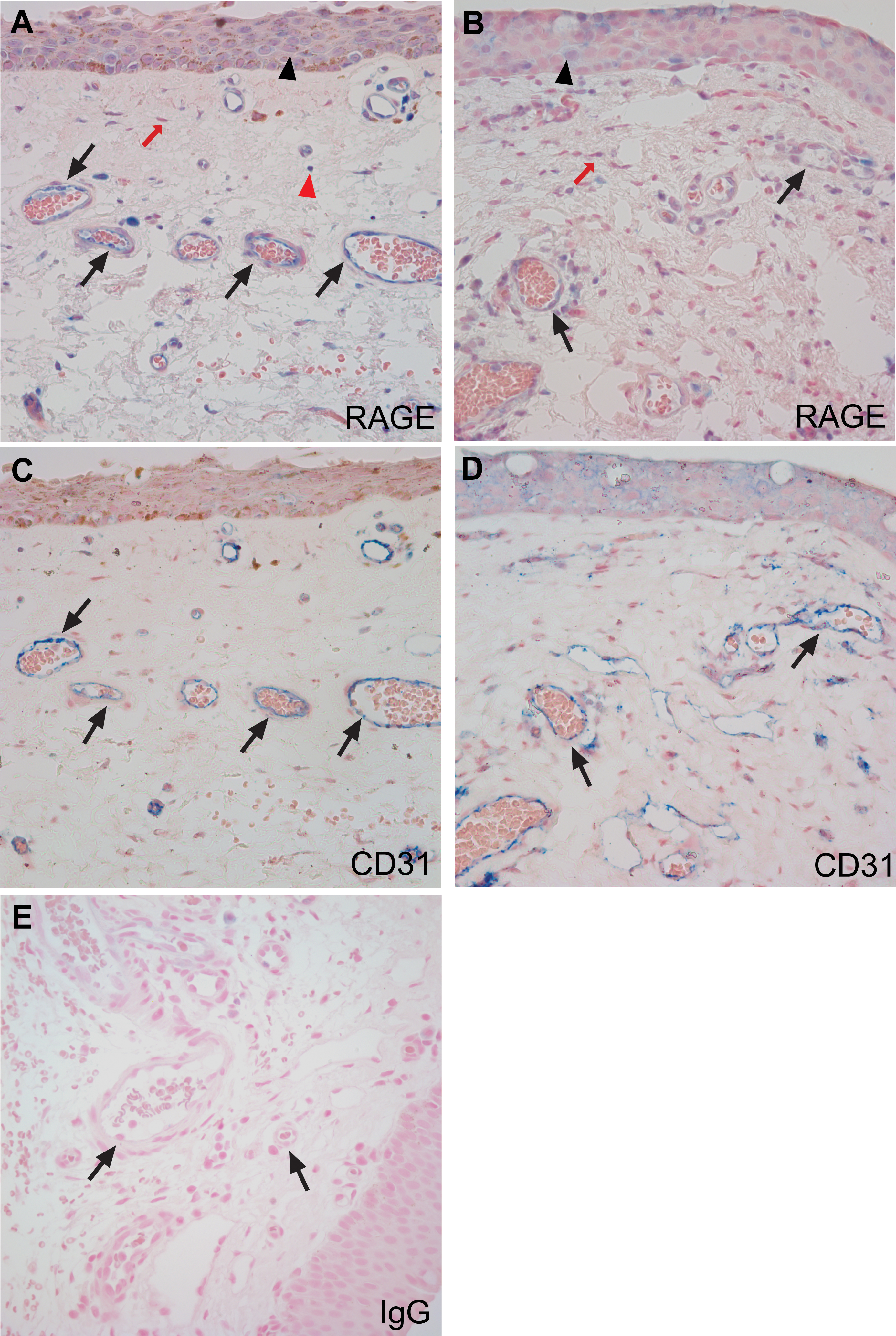
 Figure 1 of
Al-Swailem, Mol Vis 2014; 20:1740-1748.
Figure 1 of
Al-Swailem, Mol Vis 2014; 20:1740-1748.