Figure 4. Clinical case of a patient carrying an USH2A missense variants (c.1434G>C (p.Glu478Asp), c.7595–2144A>G (p.Lys2532Thrfs*56)) on one allele and an USH2A exon deletion (Del EX22) on the other allele (patient BL). A: Fundus photographs: typical form of retinitis pigmentosa. B: OCT scans: cystoid macular edema. C: Photopic electrophysiological responses: severely abnormal. D: Scotopic electrophysiological responses: severely abnormal. E: Humphrey visual field: concentrically reduced. This patient carrying a monoallelic USH2A deletion presented with the clinical picture of typical retinitis pigmentosa, similar to other cases in our series.
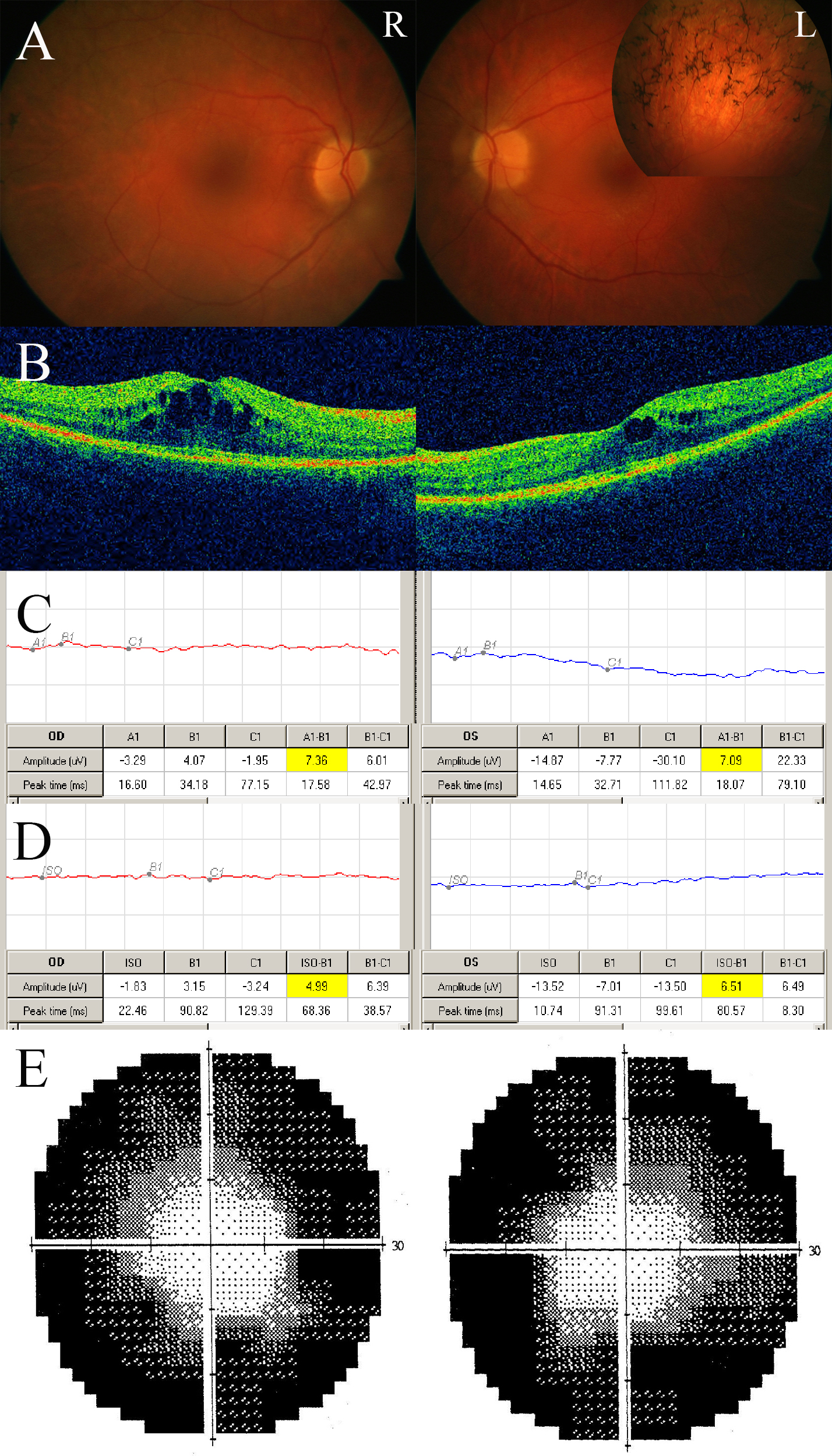
 Figure 4 of
Sodi, Mol Vis 2014; 20:1717-1731.
Figure 4 of
Sodi, Mol Vis 2014; 20:1717-1731.