Figure 1. Representative slit-lamp photographs of sub-epithelial haze and immunohistochemistry for myofibroblast marker alpha-smooth
muscle actin in rabbits. A–C: Representative slit-lamp photographs of subepithelial haze in rabbits. A: Moderate to severe (grade 3) haze (arrows) was noted in a vehicle control group cornea. B: Grade 2 subepithelial haze was observed in a rabbit eye that was treated with 0.01% resolvin E1. C: Faint subepithelial grade 0.5 haze was seen in a rabbit treated with 0.1% resolvin E1. Magnification 10X. D–F: Representative images of immunohistochemical staining for the α-smooth muscle actin (SMA) marker for myofibroblasts in the
central cornea of rabbit eyes in groups treated with (D) vehicle control solution, (E) 0.01% resolvin E1, and (F) 0.1% resolvin E1 at 4 weeks after −9D PRK. Cell nuclei are stained blue with 4',6-diamidino-2-phenylindole (DAPI) and SMA+
cells are stained red (arrows). e=epithelium. Magnification 400X.
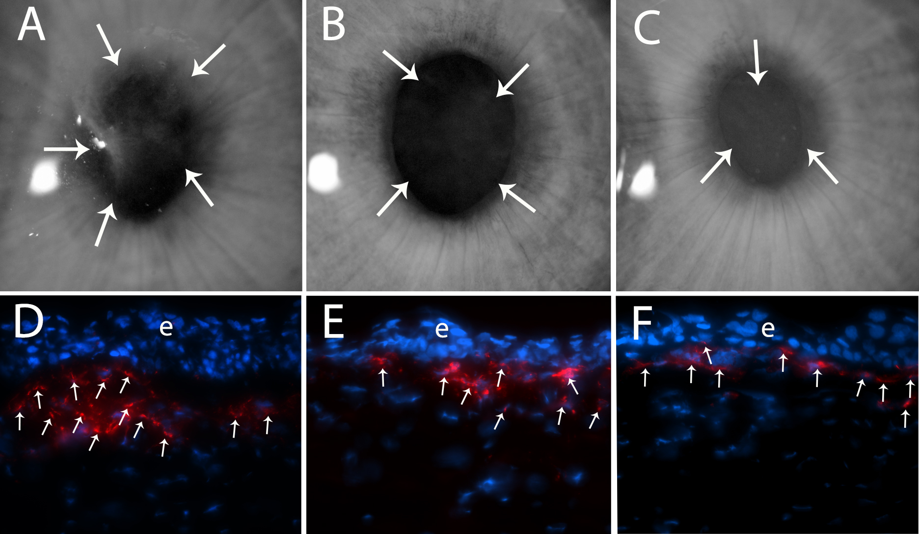
 Figure 1 of
Torricelli, Mol Vis 2014; 20:1710-1716.
Figure 1 of
Torricelli, Mol Vis 2014; 20:1710-1716.