Figure 4. Effect of echinochrome on nuclear factor κB (NFκB) p65 (red) activation in the iris/ciliary body 3 h after lipopolysaccharide
injection is shown. Dual-immunofluorescence labeling showed NFκB colocalization (yellow) in nuclei (green). A: Control naïve animals were not injected with lipopolysaccharide (LPS); an inactive NFκB signal was observed only in the
cytoplasm area of cells and no nuclear colocalization of NFκB was detected. B: The group of endotoxin-induced uveitis (EIU) rats treated with echinochrome showed reduced NFκB colocalization compared
to C: untreated EIU rats. D: Quantitative analysis of NFκB-positive cells in the iris/ciliary body (ICB). Data are shown as mean±standard error of mean
(SEM; n=4). *Amount of NFκB nuclear colocalization was significantly lower in echinochrome treated group than untreated EIU
controls (p<0.05).
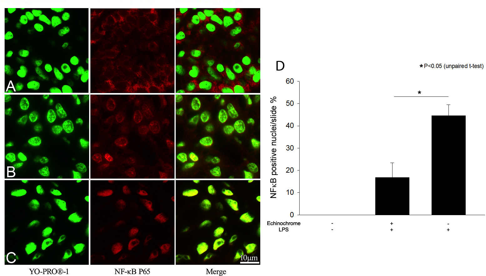
 Figure 4 of
Lennikov, Mol Vis 2014; 20:171-177.
Figure 4 of
Lennikov, Mol Vis 2014; 20:171-177.