Figure 4. Morphological assessment of iridocorneal tissues of BMP2 mouse eyes with standard histological methods. Mouse eyes were fixed,
and anterior segments (four quadrants) were processed for Epon embedding and sectioning (0.5 µm) at 7 and 36 days after Ad.cmv.BMP2
infusion. Sections were stained with 1% methylene blue, and images were captured digitally using light microscopy (top panels).
The bottom panels show the ultrastructure of the mouse outflow tissues (65 nm sections stained with uranyl acetate/lead citrate)
with transmission electron microscopy (TEM). The images are representative images of those taken from three mice in each group.
Asterisk indicates the location of Schlemm’s canal lumen; CB=ciliary body; CF=collagen fibrils; arrow points to iris synechiae
(closed angle).
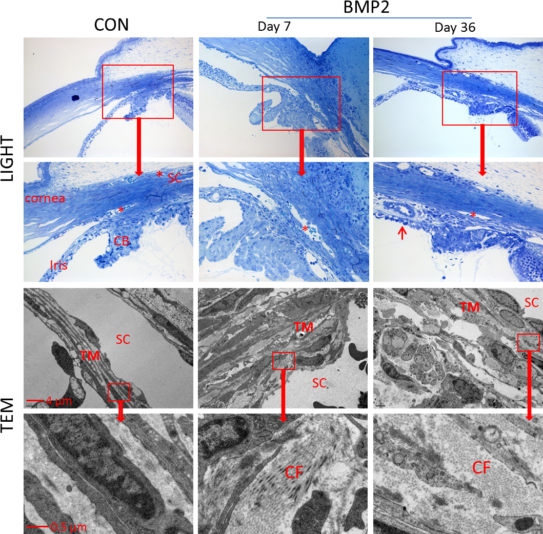
 Figure 4 of
Li, Mol Vis 2014; 20:1695-1709.
Figure 4 of
Li, Mol Vis 2014; 20:1695-1709.