Figure 3. Assessment of damage to peripheral retinal ganglion cells and optic nerves in the mouse eyes that overexpressed BMP2 in conventional
outflow tissues. Thirty-six days after the viral infusion, the mouse eyes and the optic nerves were collected. A: An image from the periphery of the flatmounted retinas of transduced mouse eyes that were immunostained with Brn3a immunoglobulin
G (IgG) and imaged with epifluorescence microscopy. B: The morphology of the myelinated axons (toluidine blue stained) of mice 36 days after intracameral transduction with BMP2.
The data are representative of images taken of four mice.
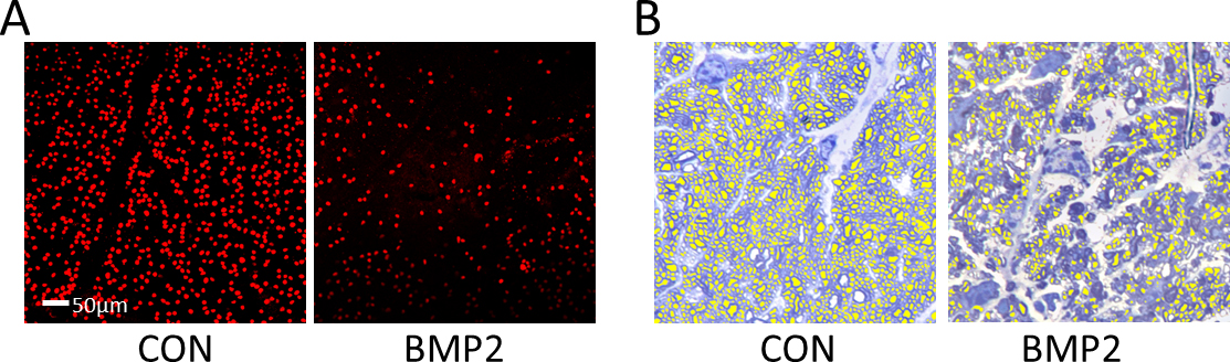
 Figure 3 of
Li, Mol Vis 2014; 20:1695-1709.
Figure 3 of
Li, Mol Vis 2014; 20:1695-1709.