Figure 1. Characterization of the BMP2 mouse glaucoma model. A: Intraocular pressure (IOP) measurements over time from mice that transiently overexpress adenovirus-mediated BMP2 protein
or green fluorescent protein (GFP) in iridocorneal angle tissues. Readings from TonoLab (T) and with direct cannulation (C)
are compared to the untreated contralateral control eyes. B: Accumulated IOP insult is compared among the three groups over the 36-day measurement period. C: The outflow facility measurements in living mouse eyes that express BMP2 or GFP to the contralateral control eyes (CON)
at 7 days post-viral infusion. D: IOP-mediated damage to retinal ganglion cells and myelinated axons in BMP2 mouse eyes at 36 days post-viral infusion. The
IOP data represent mean ± SE; n=6 for the BMP2 eyes; n=6–12 for the GFP eyes. CON is cumulative data from the contralateral
control eyes of the BMP2 and GFP infused mice (n=12–18); *p<0.05 comparing BMP2 eyes to contralateral controls; #p<0.05 comparing
BMP2 to GFP eyes. Facility and RGC/axon data are mean ± SE, n=3–4, *p<0.05 comparing BMP2 to untreated contralateral control
eyes.
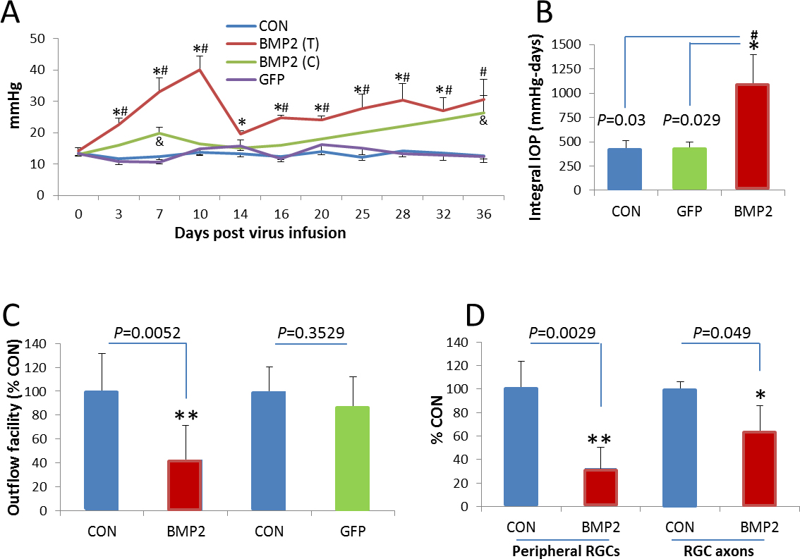
 Figure 1 of
Li, Mol Vis 2014; 20:1695-1709.
Figure 1 of
Li, Mol Vis 2014; 20:1695-1709.