Figure 3. Fluorescent antibody detection of p-Tyr-GluA2, STEP Tyr-phosphatase, PSD-95, and GABA(A)β3 receptor subunits in an approximately
4-month-old rabbit lens. The top two panels show detection of C-terminus-p-Tyr-GluA2 in the color image (left) and detection
of tyrosine phosphatase showing grayscale intensity for Alex 488 detection. The inset photo shows about 2.5-fold magnification.
The three panels below show detection of PSD-95 and GABA(A)b3 subunit in the same field in the rabbit lens (original magnification
600X and magnified about 2.5-fold).
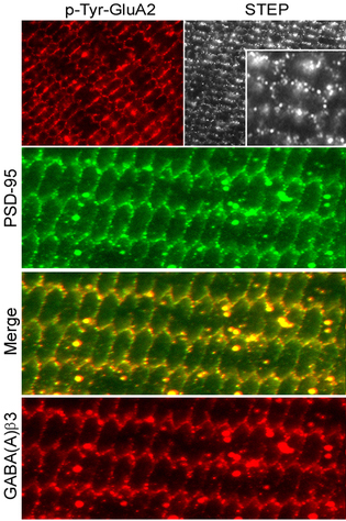
 Figure 3 of
Frederikse, Mol Vis 2014; 20:1660-1667.
Figure 3 of
Frederikse, Mol Vis 2014; 20:1660-1667.