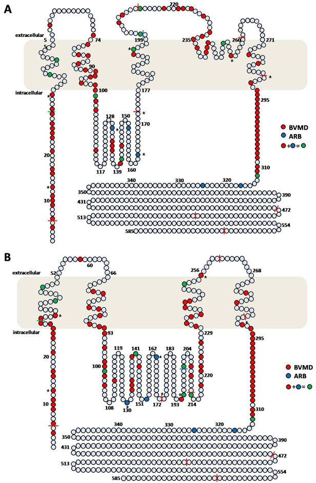
Figure 7. Diagrams of human bestrophin-1 summarizing known
BEST1 mutations associated with BVMD and ARB phenotypes [
10].
A: Protein model of bestrophin-1 proposed by Tsunenari et al. [
33];
B: Protein model of bestrophin-1 proposed by Milenkovic et al. [
34]. Colored residues indicate a missense mutation or in-frame deletion, while the colored bar indicates a nonsense or frameshift
mutation. Mutations in the homozygous or compound heterozygous state reported in the present study are marked with
*.
 Figure 7 of
Tian, Mol Vis 2014; 20:1594-1604.
Figure 7 of
Tian, Mol Vis 2014; 20:1594-1604.