Figure 6. Multiple sequenced alignment of bestrophin-1 around the mutated residues for eight species. The amino acids boxed with colors
indicate the positions of novel mutations in the present study. Mutations in a heterozygous state were marked with blue, while
mutations in a homozygous or compound heterozygous state were marked with orange. These mutations occurred in highly conserved
regions.
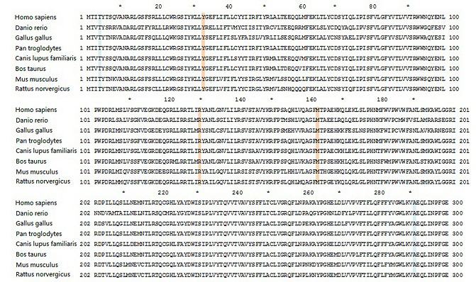
 Figure 6 of
Tian, Mol Vis 2014; 20:1594-1604.
Figure 6 of
Tian, Mol Vis 2014; 20:1594-1604.