Figure 5. Clinical evaluation of patient II:1/family H with homozygous mutation p.R130L. A, B: Fundus photography revealed a cystoid macular lesion and multiple yellowish subretinal deposits throughout the posterior
pole in both eyes. C, D: OCT showed bilateral marked intraretinal cysts in the macula and neurosensory retina detachment. OD, right eye; OS, left
eye.
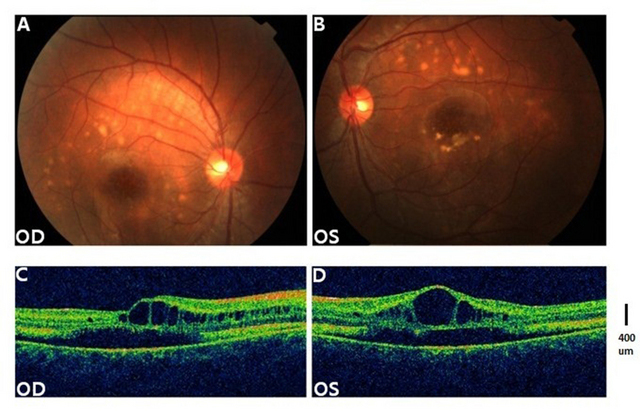
 Figure 5 of
Tian, Mol Vis 2014; 20:1594-1604.
Figure 5 of
Tian, Mol Vis 2014; 20:1594-1604.