Figure 4. Clinical evaluation of patient II:1/family G with homozygous mutation p.M163R. A, B: Fundus examination showed subretinal fibrosis in both eyes with several yellow deposits in the left eye. C, D: Optical coherence tomography (OCT) demonstrated cystoid intraretinal changes and neurosensory retina detachment in both
eyes. E, F: The fundus autofluorescence (FAF) image depicts multiple hyper-autofluorescent lesions in the peripheral retina due to deposits.
OD, right eye; OS, left eye.
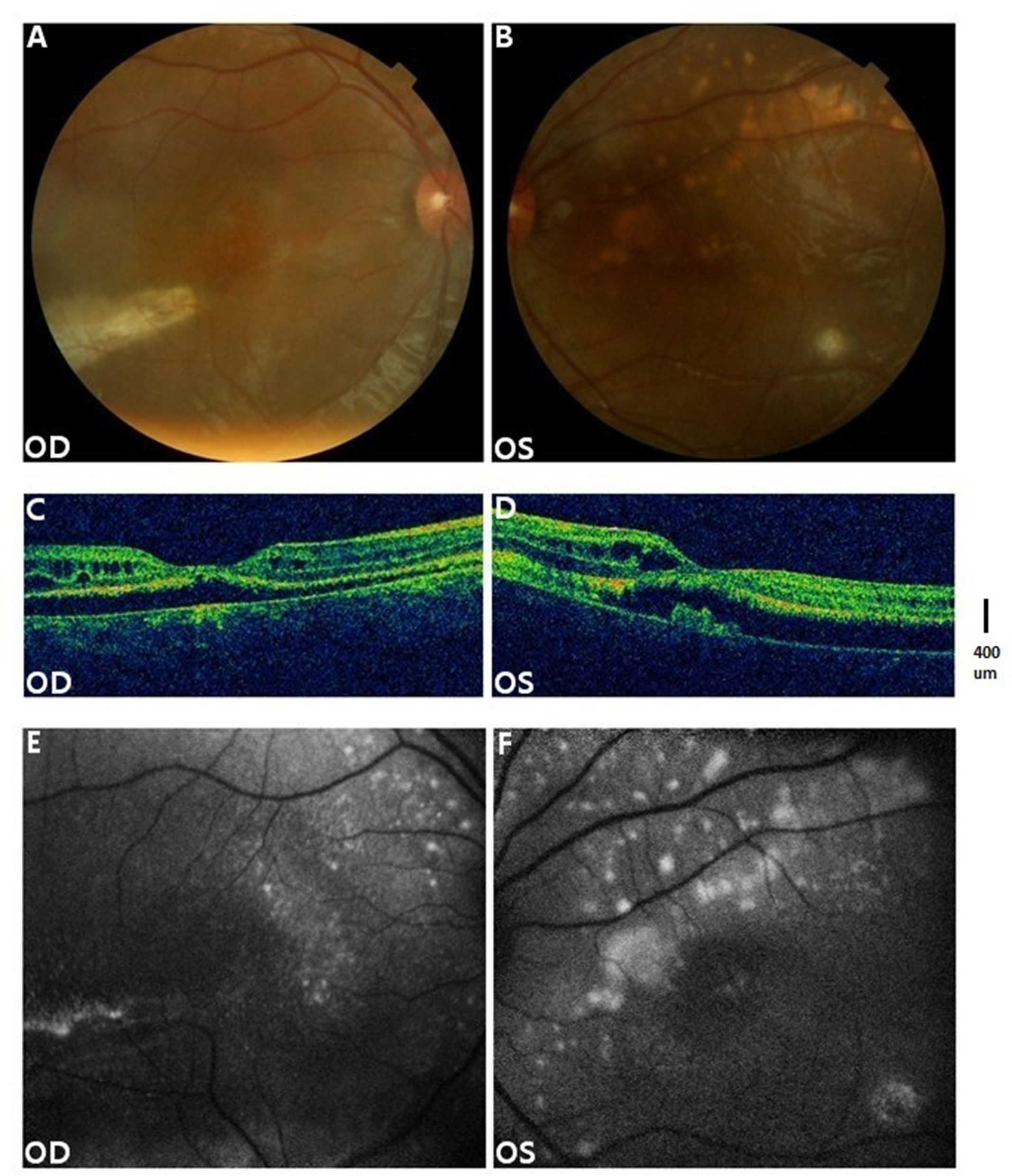
 Figure 4 of
Tian, Mol Vis 2014; 20:1594-1604.
Figure 4 of
Tian, Mol Vis 2014; 20:1594-1604.