Figure 3. Fundus photographs of patients with compound heterozygous BEST1 mutations from family I–L. A, B: Fundus photograph of patient II:1/family I reveals a macular scar in both eyes. C, D: Fundus photograph of patient II:2/family J shows a macular scar in the left eye and a vitelliruptive lesion in the right
eye. E, F: Fundus photograph of patient II:2/family K demonstrates a bilateral hyperpigmented scar. G, H: Fundus photograph of patient II:1/family L shows a pseudohypopyon lesion in the left eye and a vitelliform lesion in the
right eye. OD, right eye; OS, left eye.
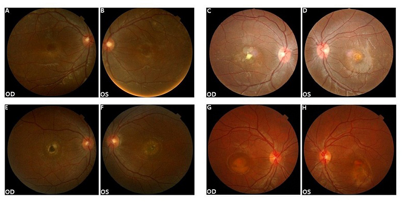
 Figure 3 of
Tian, Mol Vis 2014; 20:1594-1604.
Figure 3 of
Tian, Mol Vis 2014; 20:1594-1604.