Figure 2. Clinical evaluation of patient II:1/family B with typical BVMD. A: Fundus photograph shows a typical vitelliform lesion in both eyes. B, C: Optical coherence tomography (OCT) images demonstrate bilateral subfoveal hyper-reflective material located between the
RPE and the neuroretina. D, E: Fundus autofluorescence (FAF) images reveal marked increase in autofluorescence within the vitelliform lesion. OD, right
eye; OS, left eye.
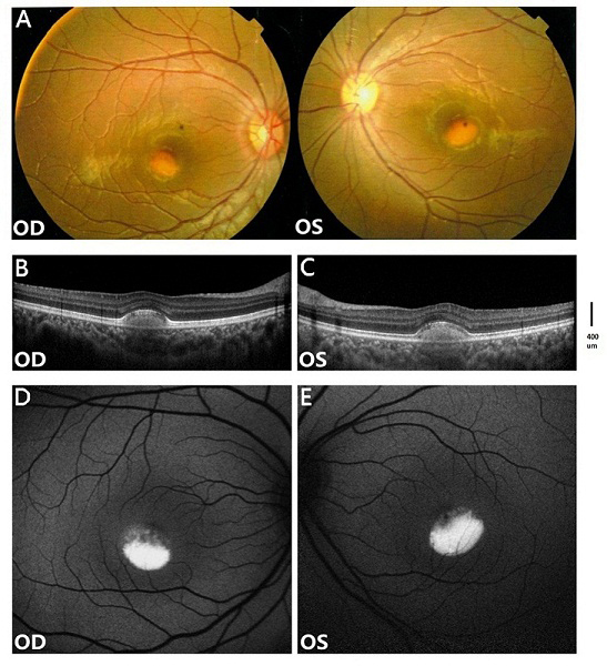
 Figure 2 of
Tian, Mol Vis 2014; 20:1594-1604.
Figure 2 of
Tian, Mol Vis 2014; 20:1594-1604.