Figure 5. Sirt1 immunohistochemistry in the aging posterior mouse eye. There were three samples (n=3) for each of the three time point,
for a total of nine animals (N=9). The representative images are shown. Sagittal sections near the optic nerve of 4- (B), 18- (D), and 24- (F) month-old mice were immunolabeled using a rabbit anti-human SIRT1 antibody that cross reacts with mouse tissue. Sirt1 labeling
was observed along the basal edge of the RPE (white arrows). Sirt1 was also detected in the OLM (black arrow), the OPL, occasional
nuclei in the INL, and the IPL. Negative controls using isotype-matched rabbit immunoglobulin (IgG) at the same concentration
as the SIRT1 antibody are shown for the 4- (A), 18- (C), and 24- (E) month-old animals. Scale bar=20 μm. OLM=outer limiting membrane, OPL=outer plexiform layer, INL=inner nuclear layer, IPL=inner
plexiform layer.
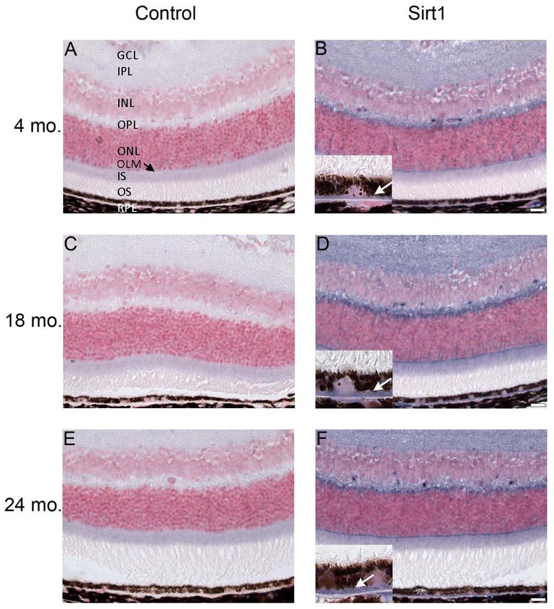
 Figure 5 of
Smit-McBride, Mol Vis 2014; 20:1569-1578.
Figure 5 of
Smit-McBride, Mol Vis 2014; 20:1569-1578.