Figure 3. Visualization of miR-34a expression in the mouse RPE using in situ hybridization. Paraffin sections of the mouse RPE were
examined using in situ hybridization at 4, 18, and 24 months of age. Images are from the ventral region near the optic nerve.
There were three samples (n=3) for each of the three time points, for a total of nine animals (N=9). The representative images
are shown. A scrambled probe was used as a negative control for each time point at 4 (A), 18 (C), and 24 months (E) of age. Negative controls did not label; pink staining is Nuclear Fast Red. A miR-34a probe was used to localize miR-34a
expression at 4 (B), 18 (D), and 24 months (F) of age. MiR-34a labeling is blue/purple (indicated with white arrows), and the nuclear counterstain is pink. At 4 months
(B), barely perceptible miR-34a expression is seen in the cytoplasm of the RPE cells. By 18 months (D), expression is much more visible and still in the cytoplasm. At 24 months (F), miR-34a expression is visibly pronounced and remains cytoplasmic. Scale bar=20 μm.
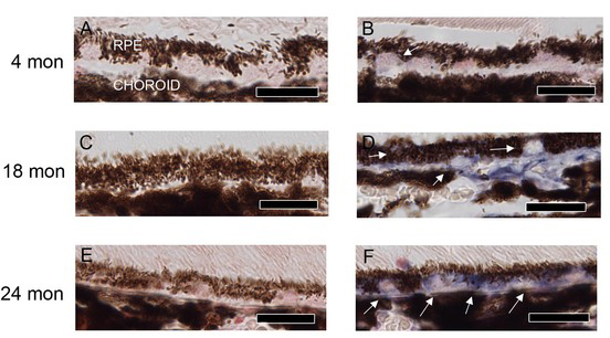
 Figure 3 of
Smit-McBride, Mol Vis 2014; 20:1569-1578.
Figure 3 of
Smit-McBride, Mol Vis 2014; 20:1569-1578.