Figure 2. Visualization of miR-34a expression in the mouse retina using in situ hybridization. Paraffin sections of the mouse retina
were examined using in situ hybridization at 4, 18, and 24 months of age. Images are from the ventral region near the optic
nerve. There were three samples (n=3) for each of the three time points, for a total of nine animals (N=9). The representative
images are shown. A scrambled probe was used as a negative control for each time point at 4 (A), 18 (C), and 24 months (E) of age. Negative controls did not label; the pink staining is Nuclear Fast Red stain. A miR-34a probe was used to localize
miR-34a expression at 4 (B), 18 (D), and 24 months (F) of age. MiR-34a labeling is blue/purple, and the nuclear counterstain is pink. At 4 months (B), miR-34a expression is localized in the inner segments (IS) and scarcely in the outer nuclear layer (ONL; see inset) of
the retina. By 18 months (D), expression is observed in the ganglion cell layer (GCL), inner nuclear layer (INL), and more intensely in the IS and the
ONL. At 24 months (F), miR-34a expression is in the GCL and the INL, with more intensity and strong expression in the IS and the ONL. Scale bar=20
μm.
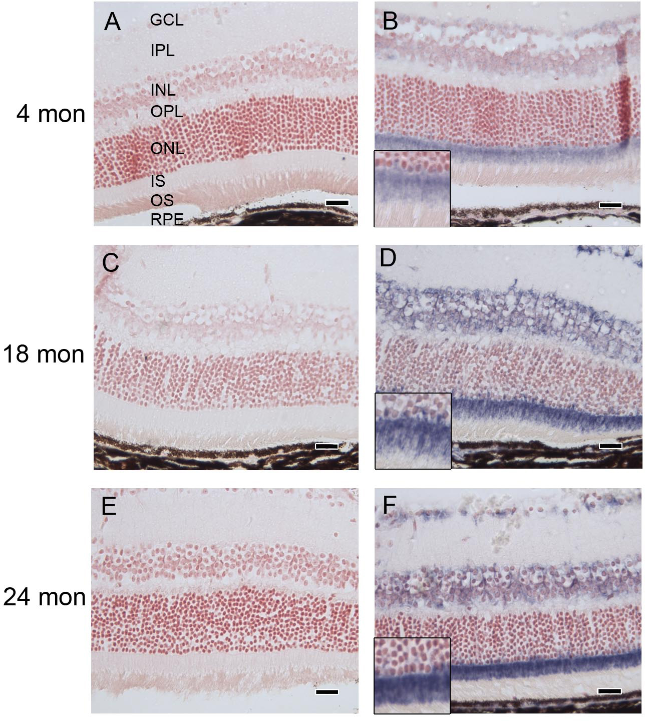
 Figure 2 of
Smit-McBride, Mol Vis 2014; 20:1569-1578.
Figure 2 of
Smit-McBride, Mol Vis 2014; 20:1569-1578.