Figure 7. Melanopsin ganglion cells in EE an ST mice. Melanopsin staining (green signal) of retinal sections from EE (A) and ST (B) rd10 mice. Red: nuclear counterstaining. Note the two plexa formed by the dendrites of the melanopsin ganglion cells at
different depths in the inner plexiform layer (IPL). Arrows point to cell bodies. INL = inner nuclear layer; GCL = ganglion
cell layer.
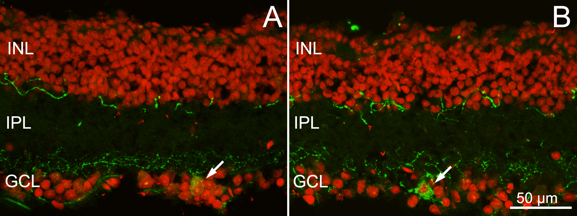
 Figure 7 of
Barone, Mol Vis 2014; 20:1545-1556.
Figure 7 of
Barone, Mol Vis 2014; 20:1545-1556.